IDEA Series: Specialised Lectures in Emergency Medicine (SLEM) – A virtual conference to strengthen EM education in the developing world

Specialised lectures in emergency medicine, virtual conference, developing world
The Problem: Emergency Medicine (EM) in Pakistan has moved from developing to developed stage in the last decade [1]. As the specialty evolves in Pakistan and other countries, there is a need to improve and assimilate novel learning methods to elevate education standards. The COVID-19 pandemic catalyzed the routine use of video-conference platforms such as Zoom. Virtual educational programming offers the opportunity to leverage educational resources across space and time, foster collaborations, and improve knowledge, clinical and evidence-based practice globally.

The Innovation

Specialised Lectures in Emergency Medicine (SLEM) is a virtual program for learning, collaboration and social engagement. The program invited experts from internationally acclaimed institutes with varying interests to present their experiences, observations, opinions, and protocols. It is an innovation that is based on a community of practice merged with the need-based assessment of a young EM residency program in a developing country.

The Learners

The target learners were EM residents and physicians practicing in the emergency department. The presenters were selected based on their experience, Free Open Access Medical (FOAM) educational materials, research, blog posts, and presentations from reputable conferences.

Group Size

SLEM accommodated 50-100 participants.

Materials

Our activity utilized simple, readily available resources. The following materials are needed:

  1. Video-conference platform: We used Zoom, a proprietary video-conferencing software program. The free plan allows up to 100 concurrent participants, with a 40-minute time restriction. Users have the option to upgrade by subscribing to a paid plan. The highest plan supports up to 1,000 concurrent participants for meetings lasting up to 30 hours. For SLEM, the paid subscription was necessary to accommodate up to 1 hour long lectures for some topics. Because of the risk of disruptive, non-invited participated, we recommend enabling the waiting room function, whereby only registered participants could join.
  2. Internet connection: A stable internet connection is a must. In order to avoid connectivity issues with Wifi, the event administrators broadcasted from an ethernet-connected computer.
  3. Engagement team: We formed a team including 5-6 residents to engage other participants and ask questions of the speakers relevant to local practice. This effort enhanced psychological safety for other participants to speak up, ask questions, and participate in the conversation following lectures.
  4. Security squad: We formed a separate team of 4 residents to oversee any non-registered participants joining the video-conference, who may generate security issues.
  5. Video library: All the lectures were recorded so that they can be referenced later by the residents.

Description of the Innovation

Speaker Identification: SLEM lecture presenters were individually approached through a defined methodology depicted in Figure 1. The program started in April 2021. The selection of the presenters was based on their published FOAM resources and scores of each were reviewed on an objective grading system that was adopted from Academic Life in Emergency Medicine (ALiEM) [2]. In addition to their content, additional factors considered included: the supporting evidence cited in their content, the referencing of their content in peer and non-peer reviewed publications, their content gradation as per the Social Media Index, and review of their faculty profiles and areas of expertise from the university website. The presenters also recommended their peer faculty who were similarly reviewed and assessed prior to the designation of the topic followed by the talk.

Topic Selection: Topics were selected based on the speaker’s previous academic lectures and area of expertise, although occasionally the presenter chose a different topic approved by the organizers based on their academic profile. Topics were selected based on disease prevalence in Pakistani EDs, published literature describing gaps in resident education and expertise, and gaps identified during academic core meetings. The presenters were then approached through either their official email address, the email address from their FOAM website, Twitter, Facebook, publications, or institution website. Upon confirmation of the lecture, an online calendar invitation including a Zoom link was shared with the presenter.

Publicity: The conference was widely advertised with promotional materials [brochure, video]via Twitter, WhatsApp, and the national EM society listserv.

Video-conference Schedule: Sessions took place virtually, starting with a 5-minute introduction of the presenter, followed by a 45-minute talk, and closing with a 15-minute question and answer session.

Lecture Evaluation: Post-session evaluation forms were shared with the residents and faculty after each session to gather feedback. Each SLEM lecture’s quality was evaluated through the internationally validated, reduced version of the Students’ Evaluation of Educational Quality (SEEQ) [3]. Originally developed by Marsh et al., this tool assesses the level of student satisfaction with teacher effectiveness to improve teaching quality. It has an excellent reliability, internal consistency, validity, and quality and has the flexibility to fit into individual teaching contexts. Reduced SEEQ is useful for quickly gathering data and decreasing the risks of item nonresponse and has been extensively studied at the postgraduate level. The variables weighted most heavily for SLEM included:

  • Learning
  • Individual rapport
  • Enthusiasm
  • Organization
  • Breadth
  • Group interaction
  • Overall rating

At the conclusion of the overall event, each participant had the opportunity to complete an online evaluation developed using Google Forms to provide feedback to the organizers. Several participants were selected for a brief, follow-up interview to explore their reactions and gain additional feedback.

The first SLEM virtual conference was successfully held July 20, 2023. Additional materials for the activity are available upon request by contacting Dr. Shahan at [email protected].

SLEM virtual conference flowchart design

Figure 1: SLEM Conference Planning and Design

Lessons Learned

SLEM has played an important role in strengthening the academic component of our developing residency. Despite the sessions being held virtually and after hours, the resident and faculty were engaged and reported increased knowledge and clinical practice improvement. Our target audience of trainees and junior to mid-level faculty especially appreciated the SLEM conference, as they appreciated tips from more senior clinicians. Additionally, the planning team developed strong bonds through the process, paving the way for future collaboration. The sessions overall contributed to the formation of a global community of practice by engaging speakers at different institutions around the world.

During planning, we faced challenges coordinating across time zones. Sending electronic calendar invitations explicitly stating the time zone along with the time was important for avoiding errors. Deploying our security teams, a robust registration system, and the waiting room function in Zoom were important strategies for avoiding disturbances to the event. Our engagement team also helped keep our participants active despite the large audience and virtual format.

IDEA series SLEM organizers

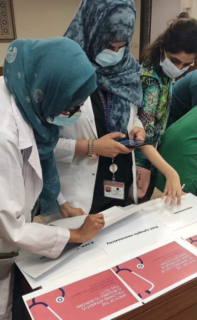
Figure 2. Team SLEM after successfully executing the SLEM conference

Theory behind the innovation

The educational theory supporting our initiative was community of practice [4]. The underlying principle highlights that learning occurs through social engagement in authentic contexts. The SLEM presenters and audiences (EM residents and faculty) were all individuals with shared interests and personal experiences relevant to the practice of EM.

Closely related, social cognitive theory also underpins the SLEM innovation. This theory postulates that learning occurs in social contexts and involves the reciprocal interaction of the individual, behavior, and the environment [5]. SLEM provided learners with the opportunity to receive experiential and tacit knowledge directly from clinical experts, which can then be applied, tested, and adjusted in their own environments. SLEM created a venue for dissemination of perspectives, discussion, and international practice change.

References

  1. Waheed S, Ali N. Chief Resident Election of Emergency Department (CREED)–An innovative approach to fair and bias-free chief resident selection in a residency program. Pakistan Journal of Medical Sciences. 2022;38(6):1717. PMID 35991269
  2. Brindley PG, Byker L, Carley S, Thoma B. Assessing on-line medical education resources: A primer for acute care medical professionals and others. Journal of the Intensive Care Society. 2022;23(3):340-4. PMID 36033246
  3. Coffey M, Gibbs G. The evaluation of the student evaluation of educational quality questionnaire (SEEQ) in UK higher education. Assessment & Evaluation in Higher Education. 2001;26(1):89-93.
  4. Schwen TM, Hara N. Community of practice: A metaphor for online design? The Information Society. 2003;19(3):257-70.
  5. Bandura A. Social cognitive theory of self-regulation. Organizational behavior and human decision processes. 1991;50(2):248-87.

IDEA Series: LUDO game to teach residents about urogenital diseases

The Problem: Urogenital diseases are commonly encountered in the emergency department [1]. Both the WHO and CDC recommend early identification and timely management of such diseases, to prevent morbidity and mortality [2, 3]. Additionally, the sensitive nature of this topic as well as cultural factors can limit the exposure and bedside teaching by emergency physicians (EPs). Novel learning methods are needed to prepare EPs to manage urogenital diseases effectively and efficiently.

The Innovation

The Learning Urogenital Diseases in Oddity (LUDO) gamification-based, timed activity teaches and assesses clinical practice essentials in the management of urogenital diseases among emergency medicine (EM) residents. This acronym plays off of the age-old game enjoyed across generations in Pakistan, also called Ludo. Adopting this popular game format with EM education enhances learning, facilitating the quick digestion of factoids [4]. The format is simple, adaptable, and can be used to teach topics in an engaging way to any learner group.

The Learners

Emergency medicine residents of all class years

IDEA LUDO game team color hats

Figure 1: LUDO game with team colors, designated by hats. Team red is listening to the question on data interpretation.

Group Size

LUDO requires a total of 3 facilitators for 4 teams, each marked with colors as per the LUDO board (green, red, blue, and yellow).

The 3 facilitator roles:

  1. One handles the laptop
  2. One marks the number of times each group has sought additional resources (limited to 5) on the whiteboard
  3. One serves as an assessor, who monitors the group whose turn it is in the game, as the throw the dice and move their color piece once they answer the question correctly.

Equipment

Figure 2: Traditional Ludo board with desk bell

Our activity utilizes simple, cheap, and readily available materials, including the following:

  • LUDO board: A square-shaped, strategy board game for 2-4 players [Ludo game on Amazon] with a pattern on it in the shape of a cross. In this game, the players race their tokens from start to finish according to the rolls of a single die.
  • LED stopwatch: An LED stopwatch was placed on the side of the projector screen. A 1-minute timer was started after the question was read to the team by the facilitator at the laptop and was reset before the next team’s turn.
  • Laptop and projector: A laptop and projector were utilized to display PowerPoint slides as a part of the activity. For example, participants had to view images, true/false questions, multiple choice questions, fill-in-the-blank questions, and rapid-fire questions on the slides. These questions prompted them to identify different aspects of urogenital diseases, as well as differentiate images of genital ulcers that patients may present at urgent care clinics or emergency departments.
  • Colored caps: Four different colored hats, matching the game pieces, were distributed to each person to designate which team they were on.
  • Desk bell: A desk bell could be rung by the team member in the “hot seat” if they wanted to answer the question differently from their team’s consensus answer.

Description of the Innovation

LUDO is a strategy board game for 2-4 players, in which teams race their 4 tokens from start to finish according to single-die rolls. A video description of the traditional Ludo game rules is below. This IDEA innovation mirrored these rules with slight modifications and took place over a 2-hour period.

Before the start of the activity, the activity director provided an introduction regarding the rules and regulations of the program that were also shared through email and WhatsApp a week before the activity.

In our LUDO variation, we assigned 4 team captains to take the “hot seats”, who were responsible for rolling the dice and had the final say in answering their team’s questions. To begin moving their tokens out of the home base circle, they must roll a 6, as in the actual game. After this, they can only move forward if they can answer the pre-prepared questions assigned to each side of the die. If they cannot answer the question, they stay where they are, and the turn proceeds to the next team. For each question, the captain has a minute to answer the question after consulting their team. An LED digital clock adjacent to the projector screen allows all participants to see the remaining time. If the captain desires to answer the question differently than the team’s consensus answer, he or she must ring the desk bell and then provide the alternative answer.

IDEA series LUDO game token movement

Figure 3: Ludo game token path – this example shows movement of the blue token

‘Getting their token home’ is the main objective of the game, but that is only part of the final evaluation to decide who is the winner. Other vital skills the assessor observes include teamwork, time management, the use of outside resources (i.e., books or the internet), and strategy. For example, there may be times when it is advantageous to answer a question wrong to increase one’s chances of landing on the other team’s token. This would send the other team’s token back to their starting position and force them to restart their travels around the board!

Each team has an option to seek additional resources to answer the question including use of our core textbook of emergency medicine or utilizing their mobile phones to access internet resources. Outside resource usage is limited 5 times per team, as tracked by a facilitator.

idea series LUDO team strategy

Figure 4: Yellow team captain joyfully removing a red team’s token piece after answer the question correctly

During the activity, the assessor facilitator judged each team’s performance through a questionnaire with Likert scales measuring the following:

  1. Knowledge of urogenital diseases
  2. Leadership skills of the team captain
  3. Problem-solving
  4. Communication among the team members
  5. Time management

At the end of the activity, each team completed an evaluation form to provide feedback about the activity to the organizers. Participants were also given timely feedback by facilitators immediately following the activity during a debriefing session, where the activity director identified gaps in knowledge and skills and provided suggestions for how to translate lessons learned to future clinical practice.

Downloadable forms:

Lessons Learned

  • The activity allowed the faculty to assess core EM skills in addition to medical knowledge, such as communication, collaboration, leadership, and problem solving. EM residents also practiced navigating team dynamics and working in a group within a safe learning space promoting collegiality among junior and senior residents.
  • The activity utilizes a popular game as a mode of introducing engagement and interest among residents and facilitates learning on a topic that does not tend to generate significant interest on its own.
  • 360-feedback was obtained:
    1. Faculty assessed learning outcomes via a Likert scale for resident feedback.
    2. Residents participated in mini-interviews at completion of the activity for educator feedback.
    3. The LUDO game’s activity and learning outcomes were assessed through a questionnaire, which was piloted prior to use during the game. The validation of the assessment tool is in process. The assessment form is available upon request by contacting Dr. Shahan.
  • The LUDO activity was well received by the EM residents, who requested to repeat the same format for other modules as well. Participants shared their feedback that this activity offers them a unique educational experience with a team-based approach.
  • Game dynamics can be altered by adjusting the complexity of the case and related questions, but any adjustments should align with the intended learning outcome. To conform to the principles of gamification, it is important that the core principles of the game should not be altered much.

Theory behind the innovation

This activity incorporated gamification and competition-based learning theory to create a positive, impactful educational experience for learners. Teaming participants in small groups facilitates collaboration and development of new knowledge through a social constructivist approach.

IDEA LUDO game

Figure 5: Happy faces at the completion of LUDO (left) and the winning group – Team Green (right)

References

By |2023-10-23T21:50:48-07:00Dec 9, 2022|IDEA series|

IDEA Series: Ultrasound-capable, 3D-printed central line trainer

Problem: Central venous line (CVL) placement is a key skill for emergency medicine providers. Sites for central line placement include the internal jugular vein, subclavian vein, and femoral vein. Indications include, but are not limited to fluid resuscitation, medication administration, central venous pressure monitoring, pulmonary artery catheter introduction, and transvenous pacing wire placement. Procedural complications can include catheter-associated infection and arterial puncture. Success rates for CVL placement vary based on location and provider experience [1-3]. Point-of-Care Ultrasound (POCUS) increases both success rate and patient safety when used to guide CVL placement [4].

central line trainer 3d idea

Figure 1. Setup for ultrasound-capable, 3D-printed central line trainer

The Innovation

The ultrasound-capable, 3D-printed central line trainer was created to facilitate realistic training of POCUS-guided CVL placement, specifically utilizing the internal jugular vein. The trainer uses a ballistic gel insert that is ultrasound-capable and replaceable, as needed.

The Learners

The model can be utilized by anyone needing practice and training on central line placement. This includes medical and physician assistant students, residents, and fellows. It will be particularly useful with students familiar with POCUS basics.

Group Size

In our experience, 4-5 students were able to utilize the model before the wear from repeated use began to impact the imaging and structure of the model, necessitating replacement of the insert. The dilation step of the Seldinger technique can be skipped or simulated in order to prolong the life of the gel insert.

Equipment

Description of the Innovation

  • The initial head model was designed using 2 common 3D modeling software systems: Tinkercad and  Meshmixer
  • A generic head and neck model was imported into Meshmixer. Using the available tools in Meshmixer, the head was rotated to the side and the neck was manipulated to enhance the appearance of an extended neck with close attention to the sternocleidomastoid muscle and clavicle.
  • The model was then imported into Tinkercad and a section of the neck was removed, inverted, and manipulated inside of a box to create a negative (mold).

central line trainer tinkercad

Figure 2. Screenshot of head being edited in Tinkercad software

central line trainer tinkercad neck

Figure 3. Screenshot of neck mold being edited in Tinkercad software

  • The head was printed with Polylactic acid (PLA) filament in 2 sections that were then glued together with superglue. The seam was sealed and smoothed with latex caulk. The files for both the head and the mold can be found in this Google Drive folder.
  • A hole was drilled from the base of the neck through the top of the head. A second hole was drilled in the base of the model.
  • To make a suitable tray for the ballistic gel insert, a thin plate was printed and then cut to fit the shape of the neck. Finally, that piece was glued to the bottom of the model.
  • The model was painted using matte spray paint.

central line trainer spray paint

Figure 4. Use of matte spray paint to paint the model

  • The mold was printed next. Two holes were drilled on either side to allow for insertion of latex tubing.
  • The ballistic gel was heated according to the directions on the box. The gel can be colored using dye or acrylic paint. Caution should be practiced when using acrylic paint. The heated gel can foam up, increasing the possibility of injury from burn.
  • While the gel was heating, the mold was prepared. The bottom was coated with a thin layer of dish soap to assist with gel release. Two sections of latex tubing, approximately 2 feet each were inserted into the mold. Modeling clay was used to fill the gaps.
  • Once colored and thoroughly heated, the gel was poured into the mold.

central line trainer mold internal jugular vein

Figure 5. Preparation of the mold in which the heated gel will be poured

central line trainer mold pour

Figure 6. The heated, colored gel is poured into the mold

  • After curing, the latex tubes were removed. The gel neck model was then removed and placed into the accompanying space on the 3d printed trainer.
  • The latex tubing was fished back through the available holes, and filled with water. As an optional step, a 30 cc syringe was attached to one end of the thicker tube. Tube stoppers can also be printed and used in place of hemostats. Pumping the syringe plunger simulates the appearance of arterial flow on ultrasound.

Video Demonstration of Final Product

Lessons learned

We are currently investigating how best to research this model. The model is inexpensive compared to available commercial CVL trainers. We estimate the cost at approximately $80 per model in materials. This, of course, does not include the price of a 3d printer, 18v drill, or drill bit. Two comparable models available for purchase are both listed for over $1000 [5, 6]. The build time is approximately 1 week with time spent printing, glue-drying, and ballistic gel setting. The model can be used repeatedly and the insert remade many times over.

If another model were to be designed, the top of the head could be sacrificed in favor of an elongated neck section. The top of the head provides no value and consumes 3d printing filament. Furthermore, an elongated neck may be preferable for a new learner by allowing more room to practice probe and hand placement.

Theory behind the innovation

Simulation as a means of teaching has been a firmly established practice across the landscape of medical education. The model in question is high-fidelity and offers the user a realistic experience in a low-stress environment. The model is small enough to be portable and can be used with little preparation, making it an ideal tool for just-in-time training in the emergency department.

Tools that allow the learner to practice multiple steps of a skill during one exercise are invaluable for skill development, competency-based medical education and mastery learning.

References

  1. McGee DC, Gould MK. Preventing complications of central venous catheterization. New England Journal of Medicine. 2003;348(12):1123-1133. doi:10.1056/nejmra011883
  2. Schummer W, Köditz JA, Schelenz C, Reinhart K, Sakka SG. Pre-procedure ultrasound increases the success and safety of central venous catheterization. British Journal of Anaesthesia. 2014;113(1):122-129. doi:10.1093/bja/aeu049
  3. E Portalatin M, Fakhoury E, Brancato R, et al. Factors contributing to unsuccessful central line placement in the neck and chest. Surgery: Current Trends and Innovations. 2019;3(2):1-5. doi:10.24966/scti-7284/100015
  4. Saugel B, Scheeren TWL, Teboul J-L. Ultrasound-guided central venous catheter placement: A Structured Review and recommendations for Clinical Practice – Critical Care. BioMed Central. Published August 28, 2017. Accessed September 21, 2022.
  5. Life/form Central Venous Cannulation Simulator. Universal Medical. . Accessed September 21, 2022.
  6. Blue Phantom internal jugular Central Line Ultrasound manikin. 3012495 – Blue Phantom – BPP-060 – Ultrasound Trainers. Accessed September 21, 2022.

IDEA | Airway Series: Reviewing Intubation Footage in Conference to Improve Airway Mastery

IDEA video airway

Airway management is one of the most critical skills learned by emergency medicine (EM) residents and can be difficult to teach in traditional lectures. Increasingly, video laryngoscopy has been utilized by emergency departments partially due to its increasing first-pass success in certain situations [1]. Additionally from a training perspective, video laryngoscopy has proven to be beneficial as attendings can have the same view as residents and provide real time feedback [2]. However, this valuable real-time feedback and anatomy visualization ability has not always been utilized in other situations such as resident conferences. In this post, we highlight how to use videos from the GlideScope (or any video laryngoscopy tool) of actual airway attempts to teach airway skills and anatomy recognition at resident educational conferences.

The Innovation

Using Video Laryngoscopy Recordings to Improve Identification of Airway Anatomy

This innovation utilizes the recording function on certain GlideScope machines to capture videos of intubations. The videos can be used during conference didactics to quiz learners on airway anatomy. They also allow learners to see a variety of difficult intubations and the different troubleshooting techniques employed.

The Learners

Although this innovation targets PGY1 EM residents, it can be beneficial to medical students or any residents struggling with intubation. It can also serve as a refresher of airway anatomy and aid in advanced troubleshooting techniques for senior residents.

Equipment

  • GlideScope machine (or any other video laryngoscopy unit) with a video record function
  • Lecture slides for material review

Description of the Innovation

During the resident didactic conference, residents, students, and attendings were presented with a series of pictures and videos recorded on a GlideScope machine, showcasing intubations performed in our ED. They then completed a timed quiz, assessing the following areas:

  • Anatomy identification
  • Airway grading
  • Identification of difficulties
  • Critique of the techniques used
  • Methods for improving the intubation

Subsequently, a brief lecture reviewing airway anatomy and intubation techniques. Finally, learners were presented with the same series of images and quizzed again.

Figure 1. Example of quiz question featuring a difficult airway image assessment question

IDEA video airway

Figure 2. Example of a match-format quiz question assessing airway anatomy

Lessons Learned

Our GlideScope machine automatically records all intubation procedures, which made putting this together easier. For institutions unable to record videos, several blogs have collections of airway footage that could be utilized, such as AirwayCam.

Quizzes were performed before and after the airway lecture. Automated grading showed significant improvement in learner performance after the lecture.
Audience feedback was overwhelmingly positive with 96% of the participants stating that the innovation would help them with real-time airway evaluation. Sample audience comments:

  • “Love this. Very educational and entertaining”
  • “Excellent session and informative”
  • “Please continue providing videos for us to learn.”

Educational Theory

This project is based on visual and applied learning. Additionally it utilizes a “learning from errors” framework which Tulis proposes can trigger emotional and motivational self regulatory learning processes [3].

Closing Thoughts

The innovation is logistically simple, is easy to replicate, received overwhelmingly positive feedback, and markedly improved scores post-lecture. Even considering the high volume of intubation experiences in the emergency department, there is always room for improvement in the learning process for this high-stakes procedure. Often we do not take the take the time to stop and critique our technique in real-time to reflect and improve. The applied and visual learning concepts serve to reinforce skills of advanced learners and build the skills of novice learners. The hope and expectation is that with serial utilization of recordings, learners will have improved recognition of difficult airways and anatomy. This in turn will hopefully lead to improving our first-pass intubation success rates.

The authors and ALiEM do not have any financial affiliations with GlideScope or any other video laryngoscopy companies.

References

  1. Brown CA 3rd, Kaji AH, Fantegrossi A, et al. Video Laryngoscopy Compared to Augmented Direct Laryngoscopy in Adult Emergency Department Tracheal Intubations: A National Emergency Airway Registry (NEAR) Study. Acad Emerg Med. 2020;27(2):100-108. PMID 31957174
  2. Monette DL, Brown CA 3rd, Benoit JL, et al. The Impact of Video Laryngoscopy on the Clinical Learning Environment of Emergency Medicine Residents: A Report of 14,313 Intubations. AEM Educ Train. 2019;3(2):156-162. Published 2019 Jan 15. PMID 31008427
  3. Tulis M, Steuer G, Dresel M. Learning from errors: A model of individual processes. Frontline Learning Research; 2016. https://doi.org/10.14786/flr.v4i2.168
By |2022-04-15T15:58:15-07:00Apr 19, 2022|IDEA series, Medical Education|

IDEA Series: Escape the Snake Room

IDEA series snake room

The Problem

A snakebite from a venomous snake can result in a potentially life-threatening toxin-mediated disease (1). The WHO considers snakebites to be an important occupational disease in Southeast Asia (2). Particularly in rural areas of Pakistan, snakebites represent a common public health concern. The relatively rare nature of this condition in urban environments, however, limits exposure to it by emergency medicine (EM) residents. Thus, additional focused training is necessary to prepare EM physicians to manage snakebites in a timely and effective manner. 

The Innovation

The “Snake Room” gamification-based, timed activity teaches and assesses clinical practice essentials in the management of snakebites among EM residents. 

The Learners

The target learners were EM residents of all class years, although a similar instructional model could be applied for teaching other uncommon diseases in under-resourced settings to any relevant learner group.

Group Size

Snake Room requires 4 total facilitators (1 facilitator per station for 4 stations). Each facilitator supervises 5-7 participants in each group during the time they attempt the station, for a total of up to 28 participants and 4 facilitators. 

Equipment

This activity utilizes simple and readily available materials, including the following:

  • WHO manual of snakebite management (3): The manual is a comprehensive guide for snakebite management specifically in Southeast Asia. The manual provides management strategies for low-resource settings of relevance to rural areas of our country. The stations were therefore developed in accordance with this reference.
  • Online stopwatch: An online stopwatch was projected on a large screen. A 15-minute timer was started at the beginning of the activity for each group, and was reset before the entry of the next group. 
  • Laptops and speakers: In 2 of the sub-stations, a computer was utilized to display PowerPoint slides as a part of the activity. For example, participants had to view images on the slides and rapidly identify different grades of snake bites, as well as differentiate images of venomous snake bites from other bites (i.e., rodent bites) that patients may present in rural areas. The slides automatically cycled after every 30 seconds. The speakers played snake charmer music in order to create an auditory distraction for the participants to mimic the distracting environment of the ED. 
  • Materials for low-fidelity wound simulation
    • Red slime to mimic features of myonecrosis 
    • Clear occlusive dressings to hold the red slime in place
    • Red, orange, and yellow dry pastel to demarcate inflammation around the bite wound
    • Manikin to demonstrate the bite marks on the lower limb

Description of the Innovation

The “Snake Room” activity took place over a 3-hour period. Four groups consisting of 5-7 members participated. There were 4 stations with 4-5 sub-stations in each station. Each station incorporated gamification and competitive-learning methodology. The substations featured clinical cases, image identification, puzzles, finding the right answer card hidden in the room, and/or low-fidelity wound simulation.

Each group of participants included residents from each class year in order to mimic the team composition most commonly encountered in our clinical environment, where senior level residents supervise a team of junior residents. The activity organizers intentionally formed teams with uniformity in academic and clinical skills to create a level playing field.

Before the start of each station, facilitators also provided a briefing to the group of participants regarding the task and amount of time available to complete it. A projector displayed the time. Groups had 15 minutes to complete each station, and each group attended the stations in the sequential order. Group members had the option to utilize online and/or in-print resources in addition to recall to complete the tasks. 

Effective use of technology was assessed. One of the groups used the Google scan app to identify the key word and obtain the answer to the puzzle.

During the activity, the course director and facilitators actively assessed participant performance as they attempted to work through the stations utilizing a questionnaire with Likert scales measuring the following:

  • Knowledge of snakebite management
  • Problem-solving
  • Leadership skills
  • Communication among team members
  • Allocation of roles among team members
  • Utilization of technology (i.e., mobile devices)
  • Understanding of the task
  • Ability to finish the activity on time
IDEA series snake room completed tasks
Successful completion of the tasks and escape from the Snake Room

The group that completed all of the puzzles and stations successfully in the allotted amount of time and achieved the highest score on the assessments of leadership, task delegation, and communication skills won the competition. 

IDEA Snake Room debrief
Debriefing session with one of the groups

At the conclusion of the activity, participants completed an evaluation form to provide feedback about the activity to the faculty organizers. Participants also received feedback during a debrief session, where faculty identified gaps in knowledge and skills and provided suggestions for how to translate lessons learned to future clinical performance. Later that day, the winning group was announced and each group’s feedback was shared in a single email to all participants, allowing groups to compare their performance. 

Materials for the activity are available upon request by contacting Dr. Shahan at [email protected].

Lessons Learned

  1. The activity allowed faculty to assess core EM skills apart from medical knowledge, such as leadership and communication. EM residents had the opportunity to practice navigating team dynamics, and working in a group within a safe learning environment facilitated collegiality among junior and senior residents.
  2. The faculty who planned the activity sought feedback from participants to optimize future iterations through short-mini interviews with group participants at the end of the activity.
  3. The assessment questionnaire was developed according to local considerations and may warrant adjustment depending on the institution and location in which the activity occurs. Internal and external validation of the assessment tool is in process. 
  4. Substations require careful, intentional planning such that they focus on an isolated aspect of the main theme, such as presentation of the disease or diagnostic test interpretation. 
  5. We incorporated audio distractions to mimic the challenges inherent to the ED environment, where residents must commonly navigate complex clinical scenarios as a team amidst frequent interruptions and competing demands. Visual distractions could also be incorporated. 
  6. Simulations (low- or high-fidelity) can be introduced while planning these sessions, but it should align with the intended learning outcomes and must be appropriately timed to maintain gamification principles. 
  7. The Snake Room activity was well received by EM residents, who requested additional iterations of this activity adapted to other topics. Participants shared their general impression that this activity offers a fun, unique educational experience with a team-based approach. 

Theory behind the innovation

We successfully combined competitive-learning theory with gamification in the Snake Room didactic to result in a positive, impactful educational experience for learners (4). Teaming participants in small groups encouraged collaboration and co-construction of new knowledge in a social constructivist approach. 

Interested in reading more innovations in education?

Read other Ideas in Didactics and Educational Activities (IDEA) series posts on ALiEM.

References

  1. Alirol E, Sharma SK, Bawaskar HS, Kuch U, Chappuis F. Snake bite in South Asia: a review. PLoS neglected tropical diseases. 2010;4(1):e603. PMID: 20126271
  2. Warrell DA. Guidelines for the management of snake-bites. Guidelines for the management of snake-bites. 2010. (https://apps.who.int/iris/handle/10665/204464
  3. World Health Organization. Guidelines for the clinical management of snake bites in the South-east Asia region. 2005.
  4. Robson K, Plangger K, Kietzmann JH, McCarthy I, Pitt L. Is it all a game? Understanding the principles of gamification. Business horizons. 2015;58(4):411-20.

IDEA Series: Acute Venous Thromboembolism (VTE) Escape Room

escape room

Adult learning theory supports medical educators in moving away from long lectures with minimal engagement from the learners [1]. Core emergency medicine (EM) topics lend themselves well to interactive methods such as gamification [2]. Puzzle-based activities can successfully facilitate team building in medical education [3].

EM residents commonly encounter acute venous thromboembolism (VTE) in the ED and must know the spectrum of presentations and approach to evaluation and treatment, including the use of risk stratification calculators.

The Innovation

  • To improve teaching of acute VTE to EM residents, we created a puzzle-based activity called “Acute VTE Escape Room.” Two teams competed against each other to solve the theme case by unlocking clues with mini puzzles, similar to the format of commercial escape rooms.

The Learners

  • As this game comprised part of the intern core curriculum, all participants were interns, with the puzzles targeted to the expected knowledge base of a PGY-1 EM resident.

Group Size

  • Group size was 4-5 learners

Equipment

  • Box with the ability to lock it
  • Numerical padlock
  • Tokens
  • Opaque envelopes
  • Laptop or tablet
  • Printed clues, questions and theme case components (Fig 1)
  • Note: If interested in obtaining printouts used in this activity, please contact Dr. Elspeth Pearce on Twitter (@ElspethKPearce)

escape room vte IDEA series

Figure 1. Game Materials

Description of the Innovation

Interns were split into 2 groups to compete against each other and race the clock to solve the case within 45 minutes. Two senior residents, one per group, assisted with the question-and-answer portion of the game. The interns had access to smartphones during the activity, and were encouraged to utilize them to access risk stratification tools during the first mini puzzle.

A theme case of obstructive shock secondary to catastrophic thrombosis of an IVC filter [4] was presented in pieces as the teams unlocked additional components of the case. The teams were given the case stem introducing the patient, chief complaint, and vital signs. They then had to unlock a box using a 4-digit passcode. This first mini puzzle had 3 cases with risk stratification scores that could be deduced. Once they calculated the risk scores they were able to unlock the box and were given the theme case history of present illness, physical exam, and instructions for the next puzzle.

The groups then had to order diagnostic laboratory and imaging tests to further evaluate the patient described in the theme case. Results were made available for tokens, with the cost of the tests similar in scale to what patients might encounter in the ED. This corresponded to an added educational objective to teach residents about resource utilization and cost of care. Labs and ECG cost 1 token and more expensive diagnostics cost 3-5 tokens. The teams earned the tokens by answering written board exam style questions (some sourced from existing board review question banks and others written by the instructor) from volunteer senior residents. Participants received the results of the tests in envelopes after they purchased them with tokens. The envelope for the lower extremity Doppler ultrasound included an additional puzzle necessitating completion in order to obtain the results.

The final mini puzzle included 4 ECGs that could be seen in acute pulmonary embolism with four questions to answer. Participants filled in the answers in boxes. Highlighted boxes yielded a passcode required to access a PowerPoint that then revealed a video of a positive ultrasound for DVT. The interns were expected to interpret this ultrasound, apply this result to the case components they had obtained, and report the final diagnosis and treatment to the instructor. A prize was awarded to the winning team.

Both groups had 45 minutes to complete the activity, allowing the instructor roughly 10 minutes to debrief, answer questions, and deliver a brief lecture on acute VTE. After completion of the activity, the participants filled out a survey evaluating the activity.

Figure 2. Learners Solving Mini Puzzle 2


Figure 3. Learners Solving Mini Puzzle 3


Outcomes

This activity was completed in-person during the hour designated for the intern core curriculum prior to the start of the resident didactic conference. Nine out of ten (90%) interns completed the acute VTE escape room and 6 (66.7%) completed the post event survey (Fig 2). Both groups finished in the time allotted with one group requiring help from the instructor to finish on time. All participants agreed or strongly agreed that the time was used effectively, and the material was presented in a clear and organized manner. Five participants (83.3%) strongly agreed that the material was delivered in an enthusiastic and stimulating way. The comments on the activity were overwhelmingly positive: “Fun and engaging way to learn about the topic”, “I LOVED this activity and really enjoyed it! Thanks for organizing it!”

IDEA Escape Room survey results

Figure 4. Survey Results

Lessons Learned

We successfully developed this game for a small group of residents at approximately the same level of medical knowledge. Adjusting the activity to target a more heterogeneous knowledge base would allow for participation by EM residents of all levels. The questions used for obtaining tokens (mini puzzle 2) and the ECG reading (mini puzzle 3) could be adjusted to the level of learner. We would recommend small group sizes as we discovered the printouts were hard to share amongst the whole group. The debrief session at the end provided a key opportunity to address any remaining questions among learners and clarify any ongoing knowledge gaps. Both groups needed some explanation of the theme case given that it involved a rare and difficult diagnosis to make. As both groups answered some of the most difficult board review questions incorrectly, future iterations may seek to better target questions to the level of the learner.

Theory Behind the Innovation

Gamification, as described by Bíró in 2014, was used as the educational theory foundation for this escape room style activity [5]. Each learner working with a team could create their own path to the correct answers. The groups and the competitive environment provided the motivation to quickly learn and adapt to the puzzles presented. 

The debrief session at the end allowed us to address existing gaps in medical knowledge and unpack emotions experienced by participants during gameplay. Debriefing theory allows the instructors of an activity, usually simulation, to create an emotionally charged event within a safe space for learning [6]. Through the debrief, instructors can identify and address gaps in clinical knowledge uncovered during gameplay.


Read other IDEA Series innovations.

References

  1. Cooper AZ, Richards JB. Lectures for Adult Learners: Breaking Old Habits in Graduate Medical Education. Am J Med. 2017 Mar;130(3):376-381. Epub 2016 Nov 28. PMID: 27908794.
  2. IDEA Series: Toxicology Virtual Escape Room during COVID-19. Academic Life in Emergency Medicine. Accessed September 22, 2021.
  3. Zhang XC, Lee H, Rodriguez C, Rudner J, Chan TM, Papanagnou D. Trapped as a Group, Escape as a Team: Applying Gamification to Incorporate Team-building Skills Through an “Escape Room” Experience. Published online 2018. doi:10.7759/cureus.2256
  4. Pearce EK. An Uncommon Cause of Shock: Acute Thrombosis of the Inferior Vena Cava. J Emerg Med. 2021 Jul;61(1):67-69. Epub 2021 May 8. PMID: 33972133.
  5. Bíró GI. Didactics 2.0: A Pedagogical Analysis of Gamification Theory from a Comparative Perspective with a Special View to the Components of Learning. Procedia – Soc Behav Sci. 2014;141:148-151. Doi: 10.1016/j.sbspro.2014.05.027
  6. Fanning RM, Gaba DM. The role of debriefing in simulation-based learning. Simul Healthc. 2007 Summer;2(2):115-25. PMID: 19088616
By |2021-11-26T16:53:23-08:00Nov 24, 2021|IDEA series, Medical Education|

IDEA Series: DIY Suture Kit Station

laceration suture repair closure

In medical training there is a lack of simulation based activities including procedural labs. Suturing is a critical skill for trainees to master in the emergency department. However, supervised practice is needed prior to suturing a real patient for the first time. This innovation allows early trainees to master suturing while on shift, using easy to find materials, which increases procedural competency and confidence. This activity allows the teacher to assess and correct the trainees procedural skills prior to attempting to suture a real patient.

Name of innovation

  • This Do-It-Yourself Suture Kit Station incorporates easy to find materials available in every emergency department, allowing early trainees to master suturing prior to suturing real patients.

Learners targeted

  • Medical students and early trainees who need suture practice

General group size

  • One-on-one student training is ideal, but can have multiple students who can practice using multiple suturing stations
  • If teacher unable to instruct while on shift, trainees can be shown a suture training video and practice alongside the video

DIY suture training kit for laceration repair

Materials needed

  • Blue chuck pad
  • Paper/cloth tape
  • Scalpel
  • Suture material
  • Suture kit

More detailed description of the activity and how it was run

  • Make the DIY Suture Kit Station (see above video):
    • Place a thick chuck pad on a flat sturdy surface.
    • Apply cloth tape to the entire surface of the chuck, and tape over the chuck. This is now the suturing pad.
    • Use a scalpel to make an incision to the pad.
    • Use the back blunt end of the scalpel to ‘fluff’ up incision edges to make laceration.
  • Use a laceration repair kit and suture to close the laceration.
  • Instruct the trainee on proper suturing technique on the suture station (or show a suture training video)
  • Have the trainee continue practicing until adequate comfort and proficiency level is achieved
  • Suture real patient!

Lessons learned, especially with regard to increasing resident and program buy in

  • Procedural skills require much repetition to gain proficiency. This is best done with video tutorials, supervision, and deliberate practice.
  • Practicing in a simulated environment greatly improves skill and confidence in real clinical practice.

Educational theory behind the innovation including specifics/styles of teaching involved

  • Simulation practice increases procedural competency.
  • Practicing on shift allows trainees to reach the number of repetitions required to gain mastery in suturing, Routt [1] showed that the number of repetitions required to gain proficiency was 41 times.
  • Competency in suturing is required even when cases are low. Wongkietachorn et al. demonstrated that tutoring suturing improves the trainees’ skillset. A practice suture kit helps improve retention for real-life scenarios [2].

Pearls

  • This DIY suture pad station technique is easily available and inexpensive.
  • To improve suturing techniques and enhance skill retention, medical students and early trainees need to learn with guided supervision on simulated task trainers.


References

  1. Routt E, Mansouri Y, de Moll EH, Bernstein DM, Bernardo SG, Levitt J. Teaching the Simple Suture to Medical Students for Long-term Retention of Skill. JAMA Dermatol. 2015 Jul;151(7):761-5. doi: 10.1001/jamadermatol.2015.118. PMID: 25785695.
  2. Wongkietkachorn A, Rhunsiri P, Boonyawong P, Lawanprasert A, Tantiphlachiva K. Tutoring Trainees to Suture: An Alternative Method for Learning How to Suture and a Way to Compensate for a Lack of Suturing Cases. J Surg Educ. 2016 May-Jun;73(3):524-8. doi: 10.1016/j.jsurg.2015.12.004. Epub 2016 Feb 20. PMID: 26907573.
By |2021-10-08T10:19:05-07:00Oct 15, 2021|IDEA series, Trauma|
Go to Top