
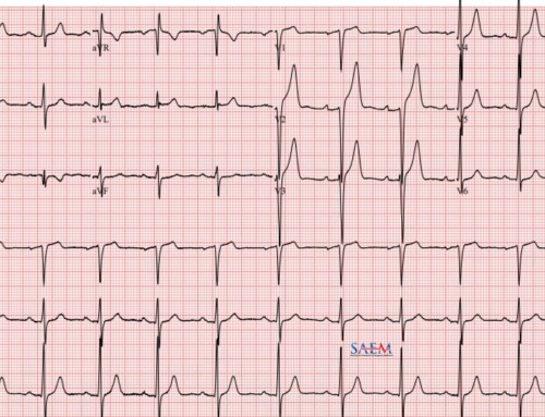
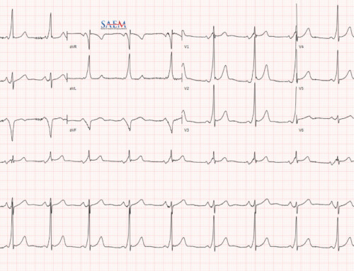
 Sudden cardiac death accounts for almost 400,000 deaths per year in the United States, and EM providers must be adept at discerning subtle, high-risk ECG findings. With the advent of triage ECG protocols, one of the most common interruptions in the ED is a request to “sign off” on an ECG. We present a reference of some of the most important high-risk ECG findings, intended to help ED providers systematically screen patients in triage and the waiting room.
Can’t Miss ECG Findings
- Did you know that there are now only 2 (not 3) Brugada ECG types?
- Did you know that the 2013 ACCF/AHA criteria for ST elevation MI1 are different from the 2012 European Society of Cardiology (ESC)? The latter also stratifies ST elevation cutoffs for men older and younger than 40 years old, and also has more specific recommendations on right sided and posterior leads.2
- Have you heard of an epsilon wave?
Use this card as a checklist to methodically ensure you are not missing any electrocardiographic evidence of red-flag conditions. [Version 2, updated July 21, 2018]
ECG tracing images were obtained from various sources:
- EKG World Encyclopedia and Wikimedia Commons [CC BY-SA 3.0 (http://creativecommons.org/licenses/by-sa/3.0)], via Wikimedia Commons, courtesy of Michael Rosengarten BEng, MD.McGill
- CardioNetworks
- Susan Lambe, MD (UCSF Emergency Medicine)
- James Heilman, MD (Own work) [CC BY-SA 3.0 (http://creativecommons.org/licenses/by-sa/3.0) or GFDL (http://www.gnu.org/copyleft/fdl.html)] via Wikimedia Commons
- W.G. de Voogt, MD, PhD, SLAZ, The Netherlands (W.G. de Voogt, MD, PhD, SLAZ, The Netherlands) [CC BY-SA 3.0 (http://creativecommons.org/licenses/by-sa/3.0)], via Wikimedia Commons (HOCM)
- By Npatchett (Own work) [CC BY-SA 4.0 (http://creativecommons.org/licenses/by-sa/4.0)], via Wikimedia Commons (second degree heart block)
References
- Moon JCC, Perez De Arenaza D, Elkington AG, et al. The Pathologic Basis of Q-Wave and Non-Q-Wave Myocardial Infarction. J. 2004;44(3):554-560. doi:10.1016/j.jacc.2004.03.076
- Gowda RM, Khan IA, Wilbur SL, Vasavada BC, Sacchi TJ. Torsade de pointes: the clinical considerations. I. 2004;96(1):1-6. doi:10.1016/j.ijcard.2003.04.055
- Lian J, Kowey PR, Yan G-X. J Wave Syndromes. In: Management of Cardiac Arrhythmias. Humana Press; 2010:453-470. doi:10.1007/978-1-60761-161-5_21
- Charlton NP, Lawrence DT, Brady WJ, Kirk MA, Holstege CP. Termination of drug-induced torsades de pointes with overdrive pacing. T. 2010;28(1):95-102. doi:10.1016/j.ajem.2008.09.029
- Antzelevitch C, Yan G-X. J wave syndromes. H. 2010;7(4):549-558. doi:10.1016/j.hrthm.2009.12.006
- Chung F, Lin Y, Chang S, et al. Current and state of the art on the electrophysiologic characteristics and catheter ablation of arrhythmogenic right ventricular dysplasia/cardiomyopathy. J Cardiol. 2015;65(6):441-450. [PubMed]
- Catanzaro J, Meraj P, Zheng S, Bloom G, Roethel M, Makaryus A. Electrocardiographic T-wave changes underlying acute cardiac and cerebral events. Am J Emerg Med. 2008;26(6):716-720. [PubMed]
- te R, Tandri H, Bluemke D. Arrhythmogenic right ventricular cardiomyopathy (ARVC): cardiovascular magnetic resonance update. J Cardiovasc Magn Reson. 2014;16:50. [PubMed]
- Fengler BT, Brady WJ, Plautz CU. Atrial fibrillation in the Wolff-Parkinson-White syndrome: ECG recognition and treatment in the ED. T. 2007;25(5):576-583. doi:10.1016/j.ajem.2006.10.017
- Pride YB, Tung P, Mohanavelu S, et al. Angiographic and Clinical Outcomes Among Patients With Acute Coronary Syndromes Presenting With Isolated Anterior ST-Segment Depression. J. 2010;3(8):806-811. doi:10.1016/j.jcin.2010.05.012
- Mattu A, Brady WJ, Robinson DA. Electrocardiographic manifestations of hyperkalemia. T. 2000;18(6):721-729. doi:10.1053/ajem.2000.7344
- Maron BJ, Ommen SR, Semsarian C, Spirito P, Olivotto I, Maron MS. Hypertrophic Cardiomyopathy. J. 2014;64(1):83-99. doi:10.1016/j.jacc.2014.05.003
- Eisenberg MJ, De Romeral LM, Heidenreich PA, Schiller NB, Evans GT Jr. The Diagnosis of Pericardial Effusion and Cardiac Tamponade by 12-Lead ECG. C. 1996;110(2):318-324. doi:10.1378/chest.110.2.318
- Punukollu G, Gowda R, Vasavada B, Khan I. Role of electrocardiography in identifying right ventricular dysfunction in acute pulmonary embolism. Am J Cardiol. 2005;96(3):450-452. [PubMed]
- O’Gara PT, Kushner FG, et al. 2013 ACCF/AHA Guideline for the Management of ST-Elevation Myocardial Infarction: A Report of the American College of Cardiology Foundation/American Heart Association Task Force on Practice Guidelines. C. 2012;127(4):e362-e425. doi:10.1161/cir.0b013e3182742cf6
- Bainey KR, Armstrong PW. Transatlantic Comparison of ST-Segment Elevation Myocardial Infarction Guidelines. J. 2016;67(2):216-229. doi:10.1016/j.jacc.2015.11.010

Christian Rose, MD
Clinical Informatics Fellow
Stanford University;
Emergency Physician, Kaiser Permanente
Stanford University;
Emergency Physician, Kaiser Permanente
Latest posts by Christian Rose, MD (see all)
- Reading from the Silver Linings Playbook: The ALiEM Connect Project - April 26, 2021
- How I Stay Healthy in EM: Christian Rose - August 21, 2020
- 9-Minute Workout for the Frontline Provider - May 15, 2020

Robert Goodnough, MD
Assistant Professor, Medical Toxicology
Department of Emergency Medicine, Baylor College of Medicine
Department of Emergency Medicine, Baylor College of Medicine
Latest posts by Robert Goodnough, MD (see all)
- ACMT Toxicology Visual Pearls: A Foraging Experience to Die For - June 8, 2020
- Can’t Miss ECG Findings for the Emergency Medicine Provider - July 11, 2018