SAEM Clinical Image Series: Pediatric Rash

pediatric rash

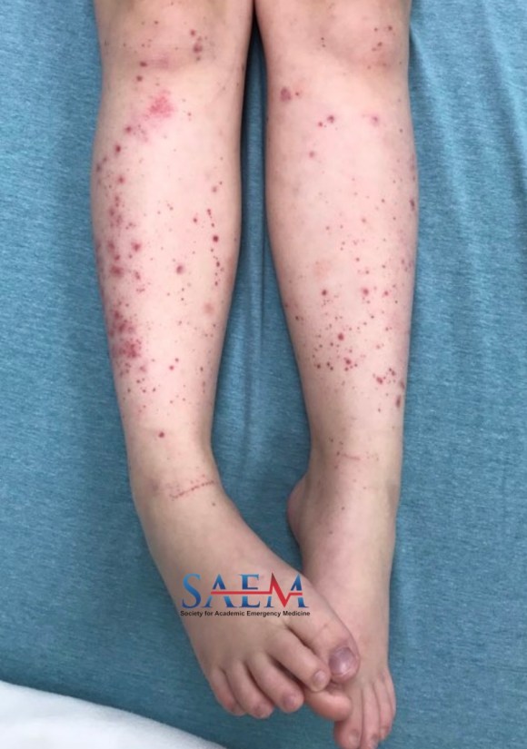
A previously healthy 8-year-old female presents to the pediatric emergency department due to a rash. Her symptoms started three days prior to presentation with a painful rash on her lower extremities. The rash subsequently spread to the buttocks and upper extremities, and she developed intermittent diffuse abdominal pain, a nonproductive cough, and pharyngitis. The patient denies subjective fever. Known sick contacts include the patient’s mother, who tested positive for COVID-19 two and a half weeks prior.


Vitals: T 98.5°F; HR 93; BP 115/68; RR 16; O2 sat 100% on room air

Constitutional: Well-developed and in no acute distress

HEENT: Normocephalic, atraumatic; moist mucus membranes; no conjunctival injection; posterior pharyngeal erythema without exudates; tonsils are three bilaterally; lips are not cracked; no “strawberry tongue”;

Neck: Normal range of motion; no lymphadenopathy

Cardiovascular: Regular rate and rhythm; normal heart sounds and pulses

Pulmonary: Effort is normal; normal breath sounds; no wheezing

Abdominal: Abdomen is flat; minimal tenderness to palpation without guarding; no organomegaly

Skin: Diffuse petechial rash and painful, palpable, nonblanching purpura in the dependent regions (most notable on the buttocks and lower extremities)

COVID-19: Detected

Complete blood count (CBC): WBC 10K, hemoglobin 13, platelets 469

Comprehensive metabolic panel (CMP): Na 138, K 4.1, Cl 103, CO2 26, BUN 7, Cr 0.38, Glucose 94, ALT 23, AST 26, Albumin 4.5

Lipase: 10

Urinalysis (UA): Normal

C-reactive protein (CRP): 3.4

Erythrocyte sedimentation rate (ESR): 24

Procalcitonin: 0.03

Fibrinogen: 363

BNP: <10

Troponin: 0.00

Ferritin: 83

Triglycerides: 37


  • COVID-19-Associated Multisystem Inflammatory Syndrome in Children (MIS-C): According to CDC criteria, patients must be under 21 years of age, with a fever higher than 38°C/subjective fever for longer than 24 hours, laboratory evidence of inflammation, severe illness requiring hospitalization, and two or more organ systems involved (cardiac, renal, respiratory, hematologic, gastrointestinal, dermatologic, or neurologic), with no alternative plausible diagnoses and recent COVID-19 infection.
  • Immunoglobulin A Vasculitis (Henoch-Schönlein Purpura): According to the EULAR/PRINTO/PRES criteria, symptoms must include cutaneous findings (palpable purpura or petechiae without the presence of thrombocytopenia), plus at least one of the following: diffuse abdominal pain with acute onset, arthritis/arthralgia, renal involvement in the form of proteinuria or hematuria, or deposition of Immunoglobulin A seen on renal histology.
  • Kawasaki Disease: The diagnostic criteria include fever for five days or longer and four of the following: bilateral conjunctival injection, cervical lymphadenopathy, polymorphous rash, oral mucous membrane changes (including fissured lips, pharyngeal erythema or strawberry tongue), peripheral extremity changes (edema of the hands/feet or desquamation).

Immunoglobulin A (IgA) Vasculitis.

This patient presented with palpable purpura and petechiae without the presence of thrombocytopenia, as well as diffuse abdominal pain. The majority of cases of IgA Vasculitis are preceded by a respiratory pathogen, with the most common being streptococcus, staphylococcus, and parainfluenza virus. Although not well-documented due to its recent conception, COVID-19 is likely to be the cause of this patient’s vasculitis. Usual management of IgA Vasculitis is supportive care, with admission and specialty referral for complications including intussusception and glomerular involvement. Given the severity of the differential diagnoses, this patient was admitted to the hospital for observation and discharged the following day with close follow-up

Take-Home Points

  • COVID-19 can cause a variety of rashes in the pediatric population, and appropriate workup including inflammatory markers, complete blood count, and comprehensive metabolic panel must be initiated to rule out severe disease. Consider obtaining a troponin, EKG, chest x-ray, echocardiogram, ferritin, prothrombin time, partial thromboplastin time, international normalized ratio, fibrinogen, urinalysis and cultures to assess for end-organ damage. If positive, and the patient appears ill, consider Multisystem Inflammatory Syndrome in Children (MIS-C).
  • IgA Vasculitis is usually caused by a respiratory pathogen. Keep it on your differential when assessing children who test positive for Covid-19.
  • Complications of IgA vasculitis include intussusception, heme-positive stool, microscopic hematuria, and periarticular disease.

  • Centers for Disease Control and Prevention, Health Alert Network. (2020). Case Definition for Multisystem Inflammatory Syndrome in Children (MIS-C). [online] Available at: https://emergency.cdc.gov/han/2020/han00432.asp.
  • Trnka P. Henoch-Schönlein purpura in children. J Paediatr Child Health. 2013 Dec;49(12):995-1003. doi: 10.1111/jpc.12403. Epub 2013 Oct 18. PMID: 24134307.
  • Ozen S, Pistorio A, Iusan SM, Bakkaloglu A, Herlin T, Brik R, Buoncompagni A, Lazar C, Bilge I, Uziel Y, Rigante D, Cantarini L, Hilario MO, Silva CA, Alegria M, Norambuena X, Belot A, Berkun Y, Estrella AI, Olivieri AN, Alpigiani MG, Rumba I, Sztajnbok F, Tambic-Bukovac L, Breda L, Al-Mayouf S, Mihaylova D, Chasnyk V, Sengler C, Klein-Gitelman M, Djeddi D, Nuno L, Pruunsild C, Brunner J, Kondi A, Pagava K, Pederzoli S, Martini A, Ruperto N; Paediatric Rheumatology International Trials Organisation (PRINTO). EULAR/PRINTO/PRES criteria for Henoch-Schönlein purpura, childhood polyarteritis nodosa, childhood Wegener granulomatosis and childhood Takayasu arteritis: Ankara 2008. Part II: Final classification criteria. Ann Rheum Dis. 2010 May;69(5):798-806. doi: 10.1136/ard.2009.116657. PMID: 20413568.
  • Royle J, Burgner D, Curtis N. The diagnosis and management of Kawasaki disease. J Paediatr Child Health. 2005 Mar;41(3):87-93. doi: 10.1111/j.1440-1754.2005.00555.x. PMID: 15790316.


Stanford’s INFODEMIC Conference on COVID-19 Misinformation: Open-access podcasts

In July 2021, Dr. Vivek Murthy became the first U.S. Surgeon General to declare health misinformation a public health crisis. Specifically, COVID-19 vaccine misinformation and disinformation on social media greatly affects vaccination rates in certain populations. Rapid increases in reliable health information about COVID-19 can be overshadowed by the spread of even greater amounts of misinformation, leading to an ‘infodemic.’

The World Health Organization defines an infodemic as:

“… too much information including false or misleading information in digital and physical environments during a disease outbreak. It causes confusion and risk-taking behaviours that can harm health. It also leads to mistrust in health authorities and undermines the public health response. An infodemic can intensify or lengthen outbreaks when people are unsure about what they need to do to protect their health and the health of people around them.”

INFODEMIC Conference on Social Media and COVID-19 Misinformation

On August 26, 2021, Stanford University’s Department of Emergency Medicine and Ethics, Society, and Technology Hub co-sponsored a unique conference to address this issue, “INFODEMIC: A Stanford Conference on Social Media and COVID-19 Misinformation.” Speakers presented virtually from around the world including experts in social media, health policy, ethics, and medicine. The conference focused on the causes of COVID-19 misinformation and mitigation strategies. Vaccine Confidence, Vaccine Hesitancy, and Vaccine Equity were among the main topics of the meeting. INFODEMIC also featured representatives from Facebook, Google, and Twitter, as well as physician influencers, to discuss the role of social media companies to address misinformation online.

Below are recordings of each of the INFODEMIC conference presentations, presented as podcasts. Video recordings of these presentations are also available to view online. The conference agenda and featured speakers are listed on the Stanford INFODEMIC website.

Podcasts

By |2021-10-06T19:40:30-07:00Oct 8, 2021|Academic, COVID19|

SAEM Clinical Image Series: Pulseless and Painful Blue Leg

painful blue leg

A 57-year-old male who works as a truck driver with a history of hypertension, type 2 diabetes, and recent COVID-19 infection presents with right lower extremity pain for two hours. He reports experiencing one day of dull aching of the right leg, then being suddenly awakened with the abrupt onset of severe right leg pain and shortness of breath. He denies chest pain. EMS reports a pulseless and painful blue leg en route. The patient denies any history of trauma, irregular heartbeat, or anticoagulation.

Vitals: T 97.5°F; BP 120/78; HR 102; RR 20; oxygen saturation 100%

General: Writhing and moaning in pain

Cardiovascular: Tachycardic; 2+ pulses in all extremities except for the right lower extremity

Pulmonary: No respiratory distress

Hemoglobin: 12.9 g/dL

INR: 1.02

Phlegmasia cerulea dolens

When a patient presents with a painful, pulseless extremity, acute limb ischemia, with etiologies including aortic dissection, arterial thromboembolism, and phlegmasia dolens, is of the highest concern. Unlike a typical deep venous thrombosis (DVT), phlegmasia dolens is a DVT that causes complete occlusion, resulting in venous congestion and hypoperfusion. Risk factors for phlegmasia dolens and DVT are the same; this patient had both a sedentary occupation and recent COVID-19 as risk factors.

Phlegmasia is usually characterized early on with pale discoloration (alba) due to patency of collaterals and later with blue/cyanotic discoloration (cerulea) after complete occlusion of the venous system. It is important to remember this exam finding might be limited in pigmented skin. Venous gangrene and compartment syndrome can be delayed exam findings. The exam should include immediate evaluation of pulses with doppler and compartment checks. Imaging modalities are controversial and should not delay vascular surgery consultation. Bedside ultrasound can be rapidly performed for clot evaluation, but CT venogram would be the preferred method for surgical planning. A common femoral vein DVT can be seen on the accompanied ultrasound and CT images. The limb should be elevated, and heparin infusion should be initiated. Surgical consultation should include a discussion of thrombectomy or catheter-directed thrombolysis.

Take-Home Points

  • Phlegmasia cerulea dolens is an uncommon complication of DVT that presents with a discolored, painful, pulseless extremity, and is associated with high morbidity and mortality.
  • Initial management includes vascular surgery consultation, elevation of the extremity, and heparinization.
  1. Baker, William, and Samuel Kim. “Risking Life And Limb: Management Of Phlegmasia AlbaAnd Cerulea Dolens”. Emra.Org, 2020, https://www.emra.org/emresident/article/risking-life-and-limb-management-of-phlegmasia–alba-and-cerulea-dolens/.
  2. Gardella L, Faulk J. Phlegmasia Alba And Cerulea Dolens. 2020 Oct 12. In: StatPearls [Internet]. Treasure Island (FL): StatPearls Publishing; 2021 Jan–. PMID: 33085284.


IDEA Series: An asynchronous EMS curriculum implemented during COVID-19

asynchronous emsThe novel coronavirus pandemic (COVID-19) resulted in the cancellation of educational experiences for emergency medicine (EM) residents at many institutions, including emergency medical services (EMS) ambulance ride alongs. The Accreditation for the Council of Graduate Medical Education (ACGME) requires that residents have educational experiences related to EMS, emergency preparedness, and disaster medicine. EMS experiences must include ground unit runs, direct medical oversight, and participation in multi-casualty incident drills [1]. There are few dedicated EMS curricula published in the literature, and those in existence incorporate physical ride-alongs [2].

(more…)

Hydroxychloroquine Toxicity

hydroxychloroquine toxicityAs the COVID-19 pandemic continues to unravel, the role of hydroxychloroquine (HCQ) in the treatment of patients with this disease has been a major focus of discussion on the news and social media. Despite the lack of good data supporting its use in the clinical setting, there have been numerous reports of individual consumption of HCQ resulting in accidental overdose and even death. It is therefore important to recognize and manage patients who may present with HCQ toxicity.

(more…)

By |2020-06-16T09:03:04-07:00Jun 17, 2020|COVID19, Tox & Medications|

Social Distancing Simulation: Tips for Leading a Virtual Session With Student Learners

We’ve all had to get a bit creative over the past few weeks. COVID-19 has ushered in an era of not only pushing healthcare workers and hospitals into uncharted territory, but also challenging the structure and delivery of medical education. Simulation education is one of many teaching modalities that is affected by this change given its case-based, in-person structure with a team of learners. These characteristics unfortunately violate the 6-foot rule of social distancing. While traditional simulation is not typically conceptualized as a virtual modality, many of its principals can be successfully adapted for remote learning.

(more…)

By |2020-05-31T19:16:34-07:00Jun 1, 2020|COVID19, Education Articles, Simulation|

I’m an Emergency Medicine Physician With COVID-19, Now What?

COVID-19 physicianA 35-year-old female emergency medicine physician presents for evaluation for severe myalgias, headache, fatigue, mild nasal congestion, profound anosmia, cough, and subjective fevers and chills. She has no measured temperature above 100.4°F, but has been taking anti-inflammatories around the clock. The day previously, she called occupational health and received testing for the novel coronavirus. The next day, her test returns positive. What happens next? We are here to share our personal experiences with COVID-19 and provide some resources to best support yourselves, your families, your learners, and your colleagues throughout this uncertain and ever-changing situation.

(more…)

By |2020-04-10T23:25:40-07:00Apr 10, 2020|COVID19, Life|
Go to Top