About Parwaiz Rashidzada, MD

Pediatric Resident
Department of Pediatrics
University of Florida College of Medicine at Jacksonville

SplintER Series: My Knee, Again!

posterior tibiofemoral dislocation knee dislocation

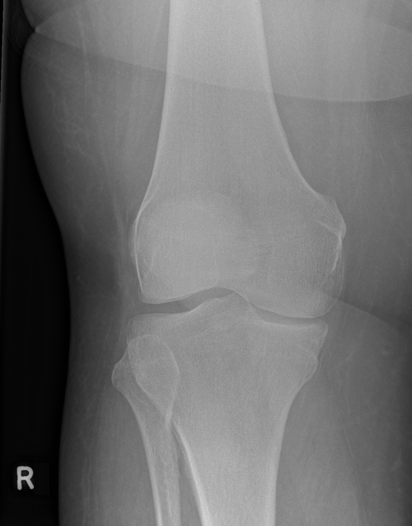
A 61-year-old F presents to the ED from the orthopedic clinic with acute right knee pain. She endorses that while a physical exam was being performed, she had sudden onset knee pain. Denies any trauma to the knee, radiation of pain, numbness, tingling, or swelling. The above knee radiographs were obtained (Images courtesy of John Kiel, DO).


(more…)

SplintER Series: I Think My Knee Popped?


patellar subluxation

13-year-old M presents to the ED with acute left knee pain that occurred about 2 hours prior to arrival while playing football. No direct trauma. Reports two audible “pops” followed by knee instability. Radiograph as pictured (Image 1. Plain film of the left knee. Image courtesy of John Kiel, DO).


Patellar subluxation. This patient likely had a spontaneous dislocation and relocation (the two “pops”). There is a very small avulsion fracture noted along the lateral femoral condyle.

  • PEARL: Patellar subluxations and dislocations are most commonly seen in the pediatric population [1].
  • PEARL: Patellar subluxation most frequently occurs in the lateral direction. Most commonly secondary to trauma, however, can also be seen in people with hypermobile joints.

It is very important to complete a full neurovascular exam. As well as performing a thorough musculoskeletal exam, assessing the anterior cruciate ligament (ACL), posterior cruciate ligament (PCL), lateral cruciate ligament (LCL), medial cruciate ligament (MCL), and patella. View the ALiEM 2 minute knee examination.

If there is an abnormal neurovascular exam or unstable knee examination, pursue further workup such as a CT scan. As an outpatient, further imaging that can be considered is an MRI knee. See below for images from this case.

Potential complications of patellar subluxations

Image 2. Knee MRI – Sagittal cut showing large knee effusion. Image courtesy of John Kiel, DO.

Potential complications of patellar subluxations

Image 3. Knee MRI – Axial cut showing the osteochondral defect of the patella. Image courtesy of John Kiel, DO.

  • PEARL: In this case, the patient has a large effusion and loose body on the outpatient MRI. The medial constraint of the patella that prevents lateral subluxation, the medial patellofemoral ligament (MFPL), is torn.

This is one of the few times a knee immobilizer is appropriate. However, close follow-up with sports medicine or orthopedics should be stressed as atrophy and contractions can occur if the patient remains in the knee immobilizer for an extended duration. Provide crutches and ask the patient to be non-weight bearing. Anti-inflammatories as needed are appropriate and encourage icing and movement.

  • PEARL: Most common complaints include pain, joint effusion/swelling, lockage, decreased range of motion, joint instability, and/or crepitation [2].

An urgent follow-up is needed with sports medicine or orthopedics for further evaluation [3]. In the case of this patient who already had an MRI, he will typically require chondroplasty of the patella and MFPL reconstruction as an outpatient. Post-operatively, he will undergo standard physical therapy with an emphasis on range of motion and quadriceps strengthening.

  • PEARL: In about 60% of the pediatric population, the zone of the MFPL injury is the predominant site of patellar insertion, which is an indication for surgical reconstruction [4].


References

  1. Chotel, F., Knorr, G., Simian, E., Dubrana, F., & Versier, G. Knee osteochondral fractures in skeletally immature patients: French multicenter study. Orthop Traumatol Surg Res. 2011;97(8). PMID: 22041573
  2. Kramer, D. E., & Pace, J. L. (2012). Acute Traumatic and Sports-Related Osteochondral Injury of the Pediatric Knee. Orthop Clin North Am. 2012;43(2), 227-236. PMID: 22480471
  3. Griffin, J. W., Gilmore, C. J., & Miller, M. D. (2013). Treatment of a Patellar Chondral Defect Using Juvenile Articular Cartilage Allograft Implantation. Arthrosc Tech. 2013;2(4). PMID: 24400181
  4. Dixit, S., & Deu, R. S. Nonoperative Treatment of Patellar Instability. Sports Med Arthrosc Rev. 2017;25(2), 72-77. PMID: 28459749


SplintER Series: A Case of Inability to Move the Knee

Normal knee radiograph

29-year-old F presents to ED with acute onset knee pain. Reports hearing an audible “pop” after twisting her leg while running down the stairs at home. She explains that her right knee is stuck, and she can neither flex nor extend it. An image is shown below (courtesy of Andrew Murphy, Radiopaedia.org)


(more…)

Go to Top