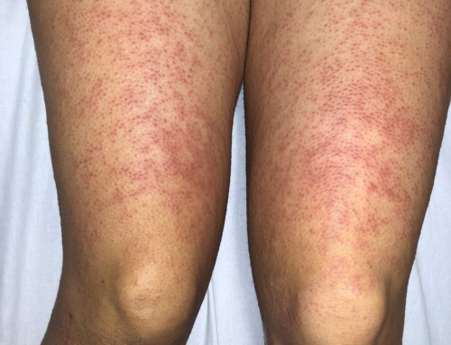
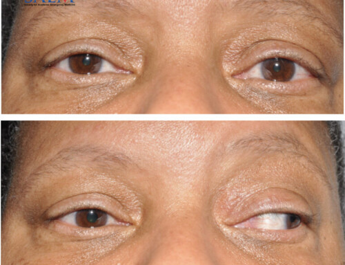
A 68-year-old male with a past medical history of hypertension, hyperlipidemia, and recent ileostomy secondary to small bowel obstruction presented for acute left arm swelling, discoloration, and numbness since last night. He endorses sudden onset of painful edema with the development of purple discoloration. He denies trauma, history of similar problems, chest pain, or shortness of breath. He endorses difficulty flexing at the elbow secondary to the amount of swelling, pain, and numbness to the arm. The patient had a peripherally inserted central catheter (PICC) line placed in the left upper extremity two weeks ago. Vitals: T 37.1°C; HR 80; BP 154/82; RR 18; O2 sat 100% on RA General: Moderate distress secondary to pain but non-toxic appearing Cardiovascular: Regular rate and rhythm; no murmurs; left ulnar artery 2+; left radial artery 1+ to palpation; bedside doppler—triphasic left ulnar artery and biphasic left radial artery; capillary refill three seconds Respiratory: Lungs clear to auscultation bilaterally; no adventitious breath sounds Musculoskeletal: Left upper extremity with global nonpitting edema from fingers to shoulder; skin with purple cyanotic discoloration; moderately tender to palpation throughout the entire limb; no crepitus or bullae; pain is not out of proportion; soft compartments throughout the left upper extremity Neurologic: Alert and oriented to person, time, and place; Glasgow Coma Scale 15; cranial nerves II-XII grossly intact; sensation decreased in left upper extremity; all other extremities intact Complete blood count (CBC): Unremarkable Partial thromboplastin time (PTT) and International normalized ratio (INR): Unremarkable Phlegmasia cerulea dolens (PCD) of the Upper Extremity. It’s just a deep venous thrombosis (DVT) right? PCD is not just another DVT, it’s a severe limb-threatening (12-25% amputation rate) and life-threatening (25-40% mortality rate) disease that presents with marked swelling in the extremity, pain, and cyanosis. The pathophysiology of PCD involves complete obstruction of both superficial and deep venous return, resulting in increased interstitial tissue pressure, arrest of capillary flow, tissue ischemia, and ultimately, gangrene. Upper extremity involvement is rare and only occurs in approximately 2-5% of all phlegmasia cases. PCD presents with key characteristics: marked edema, severe pain, pathognomonic blue discoloration/cyanosis, and eventually ischemia. Ultrasound is the best initial modality for suspected PCD and bedside ultrasound with two-point compression can be quickly performed by the emergency physician. Management should include fluid resuscitation, systemic anticoagulation, and emergent vascular surgery or interventional radiology consult for possible thrombectomy or catheter-directed thrombolysis. Images and cases from the Society of Academic Emergency Medicine (SAEM) Clinical Images Exhibit at the 2021 SAEM Annual Meeting | Copyrighted by SAEM 2021 – all rights reserved. View other cases from this Clinical Image Series on ALiEM.
Take-Home Points
Copyright

Roger Farney, DO

Latest posts by Roger Farney, DO (see all)
- SAEM Clinical Image Series: Painful Blue Arm - January 10, 2022

Branden Skarpiak, MD, DTM&H

Latest posts by Branden Skarpiak, MD, DTM&H (see all)
- SAEM Clinical Image Series: Painful Blue Arm - January 10, 2022


