

A 25-year-old medical student comes in with a muffled voice, sore throat and trismus. You look at the back of her throat and you see the uvula deviated to the right. You astutely diagnosed a peritonsillar abscess (PTA). You consider aspirating and want to check for tips on how to successfully do this.
Dr. Michelle Lin and Dr. Demian Szyld have created great guides for the common and important emergency medicine procedure of draining a PTA (laryngoscope lighting and spinal needle for aspiration; ultrasound localization and spinal needle guard; avoiding awkward one-handed needle aspiration). This update reviews these tricks as well as some additional techniques for optimal success in draining a PTA, while avoiding the ultimate feared complication of puncturing the carotid artery.
1. Use point of care ultrasonography (POCUS)
Using the intracavitary probe provides great views of both the abscess and the nearby vessels (figure 1). Not only does it confirm the presence of abscess vs. a phlegmon, POCUS also gives the operator an idea of the depth of the carotid artery with respect to the visible surface of the posterior pharynx and the TPA.

2. Visualization
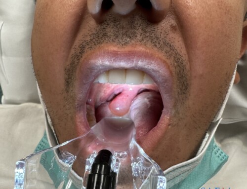
Use the bottom part of a disposable light-affixed pelvic speculum with the top removed (figure 2), leaving just one piece that the patient can hold down.
Equipment:
- Visualization tool:
- OPB Medical ER-SPEC (Figure 2), or
- C-MAC S video laryngoscope, or
- Macintosh laryngoscope blade
- Suctioning: Yankauer suction device

Pro tip #1: Note that by asking the patient to hold the light, the operator frees an extra hand for suction or stability of the other hand. Take the extra step setting up with sufficient lighting to optimize success.

3. Analgesia
Good analgesia is important for this anxiety-provoking and painful procedure. We have seen success with first using topical benzocaine spray. Alternatively, nebulizing lidocaine (2% or 4% without epinephrine) over 5 minutes can be used. Next, inject 2-3 mL of 1% lidocaine with epinephrine into the mucosa and muscle. 1
Equipment:
- Topical anesthetic spray such as HurriCaine Benzocaine
- Direct anesthetic infiltration:
- 5-10 mL syringe
- 25-27g needle to inject lidocaine
Pro tip #2: We expect the patient to salivate, or for pus to drain upon fenestration. Hand the patient the suction. Just like in the dental office, the patient can spit into the suction to optimize the view of the posterior pharynx, while minimizing risk for aspiration.
Pro tip #3: Communication is key. As it will be difficult for the patient to articulate concerns while you have the needle in hand and speculum retracting the tongue down, instruct the patient to lift the suction up to get your attention instead of saying something, or worse, pulling away. This ensures safety for the operator and the patient.
4. Drainage: Pediatric bullet tube as a depth guard
Many techniques exist for the actual drainage, including using a spinal needle or a traditional 1-1.5 inch needle, cutting the needle cover to buffer the depth of the actual needle. This protects the needle from penetrating the mucosa too deeply. Notice that the edge of the needle cover can be sharp and jagged. There is also a possibility for the plastic to chip off, thereby causing aspiration (Figure 4).

Pro tip #4. We propose an alternative approach to a trimmed needle cover sheath. Cut the top lid off a pediatric bullet tube/microtainer and insert the 18g needle through the top onto the center part of the bottom aspect of the microtainer (figure 5). 1 cm of the needle will jut out of the back end of the tube. The cut bullet tube does not leave any sharp edges. The smooth surface of the back end of this microtainer, which will avoid traumatizing the posterior pharynx upon contact, or cause pieces of plastic to dislodge.
Equipment:
- Barrier/cap: Microtainer pediatric bullet tube
- Aspiration setup:
- 5-10 mL syringe
- 18g needle

5. Aspiration
Aim the needle towards the area of most fluctuance.
Pro tip #5: Start with a 5-10 mL syringe instead of 20-30 mL syringe. This is because the larger the caliber of the syringe, the higher tension required to overcome the fluid pressure (remember LaPlace’s Law! Tension = pressure x radius), which may not be necessary in the setting of a fairly small abscess. Also, the smaller the syringe, the better access you have to the back of the patient’s throat.

Pro tip #6: While precision is key and aimed at the point of maximal fluctuance, multiple deliberate attempts actually leads to fenestrations that further allow for the pus to continue to drain post-procedure.
Microskill: Single-handed needle aspiration technique (figure 7)
- Using the operator’s dominant hand, grab the syringe with between the 1st and 2nd digits. As soon as the needle enters the mucosa, wrap the third digit around the plunger and apply negative pressure/pull back while advancing the syringe.

6. Incision
Consider performing an incision and drainage (I&D) even after aspiration with the needle and syringe for large/loculated abscesses. A 2016 Cochrane review of I&D vs. needle aspiration found low quality evidence that I&D may have lower rates of recurrence than needle aspiration.2 We have found in practice that aspiration is great for removing pus but for large or loculated abscesses a single incision with 11-blade scalpel after the aspiration allows any remaining pus to drain and thus avoid recurrence.
Pro tip #7: Tape the cover of the scalpel/retractor to the desired depth. This ensures the scalpel will not penetrate more than 1 cm. In a single deliberate approach, incise about 1 cm along the area of maximal fluctuance (figure 8).

Pro tip #8: Give the patient an ice cold water to swish and spit. It further helps control the pain, and allows for drainage to continue.
Pro tip #9: Put gloves on your patient’s dominant hand. The patient’s non-dominant hand is already holding the Yankauer suction (Figure 2). Instruct the patient to reach the newly drained abscess with their index finger, and ask the patient to place gentle pressure, “milking” the remainder fluid collection. The benefit of the patient doing this is:
• It doesn’t trigger their gag reflex as much as it would if the operator does this.
• The patient is less likely to bite their own hand.
• The pressure placed on the back actually will help alleviate some of the pain they may be experiencing.
7. Procedural success
Successful drainage is seen when:
- Fluctuance is less.
- Patient’s trismus significantly improves.
- Patient subjectively feels much better.
8. Post I&D care
PTA drainage leads to decreased oral intake, so most of these patients will be dehydrated. IV hydration may help with the patient subjectively feeling better. Oral antibiotics are always given, targeting Group A strep and oral anaerobes for 14 days.3 We suggest either regimen:
- Amoxicillin-clavulanate 875 mg PO twice daily (45 mg/kg/dose for children up to maximum of 875 mg per dose), or
- Clindamycin 300-450 mg PO every 6 hours (10 mg/kg/dose every 8 hours for children up to a maximum 450 mg per dose, although DynaMed/UpToDate states that 600 mg per dose is allowed), if MRSA is suspected.
Because of new emergence of antibiotic resistance, a culture from the abscess may be collected.4 Steroids also helps with alleviating trismus and can also reduce the pain and time of recovery.5 We suggest the regimen:
- Dexamethasone 10 mg IV or PO
Case Conclusion
The patient feels significantly better. The muffled voice improved along with the trismus. The patient is given return precautions, emphasizing symptoms such as difficulty breathing, worsening pain or trismus, difficulty tolerating oral hydration, enlarging mass, fever, neck stiffness, or bleeding. The patient is discharged home with the antibiotic regimen above plus a recommendation for over-the-counter pain medications and instruction to gargle salt water.6
References
-
Riviello R. Otolaryngologic Procedures. In: Roberts and Hedges’ Clinical Procedures in Emergency Medicine. Elsevier; 2014:1303-1309.
-
Chang B, Thamboo A, Burton M, Diamond C, Nunez D. Needle aspiration versus incision and drainage for the treatment of peritonsillar abscess. Cochrane Database Syst Rev. 2016;12:CD006287. https://www.ncbi.nlm.nih.gov/pubmed/28009937.
-
Kieff D, Bhattacharyya N, Siegel N, Salman S. Selection of antibiotics after incision and drainage of peritonsillar abscesses. Otolaryngol Head Neck Surg. 1999;120(1):57-61. https://www.ncbi.nlm.nih.gov/pubmed/9914550.
-
Sowerby L, Hussain Z, Husein M. The epidemiology, antibiotic resistance and post-discharge course of peritonsillar abscesses in London, Ontario. J Otolaryngol Head Neck Surg. 2013;42:5. https://www.ncbi.nlm.nih.gov/pubmed/23663820.
-
Chau J, Seikaly H, Harris J, Villa-Roel C, Brick C, Rowe B. Corticosteroids in peritonsillar abscess treatment: a blinded placebo-controlled clinical trial. Laryngoscope. 2014;124(1):97-103. https://www.ncbi.nlm.nih.gov/pubmed/23794382.
-
Lamkin R, Portt J. An outpatient medical treatment protocol for peritonsillar abscess. Ear Nose Throat J. 2006;85(10):658, 660. https://www.ncbi.nlm.nih.gov/pubmed/17124937.

Margaret Davis
Tufts University School of Medicine
Latest posts by Margaret Davis (see all)

Al'ai Alvarez, MD
Clinical Associate Professor
Director of Well-being
Co-Chair, The Human Potential Team
Department of Emergency Medicine
Stanford University School of Medicine
Latest posts by Al'ai Alvarez, MD (see all)
- Trick of the Trade: Managing Epistaxis with Merocel Nasal Packing and an Angiocatheter - November 11, 2022
- How I Work Smarter: Al’ai Alvarez MD - January 21, 2022
- Trick of the Trade: Persistent Paracentesis Leakage 2.0 - October 20, 2021