

The patient is a 72-year-old male with a history of CAD, hypertension, and BPH who presents to the Emergency Department for sinus congestion and right-sided facial pain. The patient reports progressively worsening darkening crusting around his nose for 3 weeks. He has also had a right-sided temporal and retrobulbar headache, blurry vision in right eye, diminished sense of smell, and right sided numbness to the roof of his mouth for the past week. He was prescribed amoxicillin and nasal steroid spray four days ago without improvement. He denies any recent illness, hospitalizations, travel, HIV risk factors, or any other complaints at this time.


Vitals: All vital signs are normal
General: Alert and oriented, speaking in clear sentences.
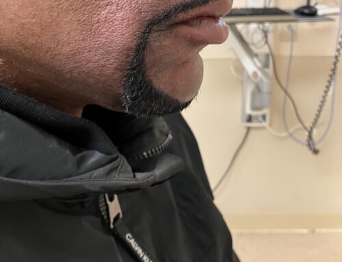
HEENT: Key findings as shown in the images provided. There is a 3cm area of palpable edema with tenderness over right temporal region. Dentition is poor with missing teeth. Tongue exam normal.
Cardiovascular: Regular rate and rhythm without murmurs.
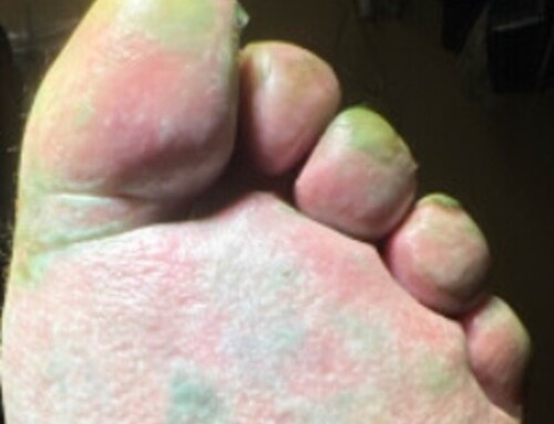
Skin: Other than as shown, no rashes
CBC: WBC 8.0, Hgb 14.9, Plt 324,000
CMP: Within normal limits
Lactate: 1.1
The patient has mucormycosis.
CT scan shows subperiosteal swelling and fluid collection measuring approximately 3.8 x 1.4 cm with a focus of gas.
One of the biggest challenges in diagnosing mucormycosis early is the nonspecific nature of its symptoms, which often overlap with more common and more benign infections. Symptoms such as fever, facial pain, and swelling are often mistaken for bacterial sinusitis. Furthermore, the rapid progression of mucormycosis means that by the time definitive diagnostic tests are conducted, the disease may have already spread significantly. Mucormycosis spreads particularly rapidly in patients with poorly controlled diabetes, neutropenia, and other immunosuppressive conditions. Definitive diagnosis relies on tissue biopsy, imaging, and molecular diagnostic methods. CT scans may show tissue necrosis, bony destruction, and soft tissue swelling in the sinuses. If rhinocerebral mucormycosis is suspected, MRI of the orbits, sinuses, and brain can evaluate for spread of infection and detect intracranial extension. Treatment includes aggressive surgical debridement of necrotic tissue and systemic anti-fungal medications.
Take-Home Points
- Mucormycosis should be suspected in rapidly progressive sinusitis with necrotic tissue or eschars around the nasal cavity or palate.
- Patients who are at high risk include those with uncontrolled diabetes or other immunocompromising conditions.
- Biopsy is gold standard for a definitive diagnosis.
- Mohamed MS, Abdel-Motaleb HY, Mobarak FA. Management of rhino-orbital mucormycosis. Saudi Medical Journal. 2015;36(7):865-868. doi:10.15537/smj.2015.7.11859.
- Gupta MK, Kumar N, Dhameja N, Sharma A, Tilak R. Laboratory diagnosis of mucormycosis: Present perspective. J Family Med Prim Care. 2022 May;11(5):1664-1671. doi: 10.4103/jfmpc.jfmpc_1479_21. Epub 2022 May 14. PMID: 35800582; PMCID: PMC9254769.
- Gamaletsou MN, McGinnis MR, Hayden RT, Kontoyiannis DP. Early clinical and laboratory diagnosis of invasive pulmonary, extrapulmonary, and disseminated mucormycosis (zygomycosis). Clin Infect Dis. 2012 Feb;54 Suppl 1:S55-60. doi: 10.1093/cid/cir868. PMID: 22247446.

Alexandria Lauray, MD
The Brooklyn Hospital Center
Latest posts by Alexandria Lauray, MD (see all)
- SAEM Clinical Images Series: No, I Am Not Diabetic! - December 5, 2025

Michael Levine, MD CHSE
Director of ED Simulation & Core Faculty
The Brooklyn Hospital Center
Latest posts by Michael Levine, MD CHSE (see all)
- SAEM Clinical Images Series: No, I Am Not Diabetic! - December 5, 2025
