ALiEM AIR Series | Orthopedics Lower Extremity Module

Welcome to the AIR Orthopedics Lower Extremity Module! After carefully reviewing all relevant posts in the past 12 months from the top 50 sites of the Social Media Index, the ALiEM AIR Team is proud to present the highest quality online content related to related to neurologic emergencies in the Emergency Department. 4 blog posts met our standard of online excellence and were approved for residency training by the AIR Series Board. More specifically, we identified 1 AIR and 3 Honorable Mentions. We recommend programs give 2 hours of III credit for this module.
AIR Stamp of Approval and Honorable Mentions
In an effort to truly emphasize the highest quality posts, we have 2 subsets of recommended resources. The AIR stamp of approval is awarded only to posts scoring above a strict scoring cut-off of ≥30 points (out of 35 total), based on our scoring instrument. The other subset is for "Honorable Mention" posts. These posts have been flagged by and agreed upon by AIR Board members as worthwhile, accurate, unbiased, and appropriately referenced despite an average score.
Take the AIR Orthopedics Lower Extremity Module at ALiEMU
Interested in taking the AIR quiz for fun or asynchronous (Individualized Interactive Instruction) credit? Please go to the above link. You will need to create a free, 1-time login account.
Highlighted Quality Posts: Orthopedic Lower Extremity Emergencies
| Site | Article | Author | Date | Label |
|---|---|---|---|---|
| PedEM Morsels | Lisfranc Injuries in Pediatric Patients | Christyn Magill, MD | 3/18/22 | AIR |
| Rebel EM | Compartment Syndrome | Anand Swaminathan, MD | 5/4/22 | HM |
| EM Cases | Emergency Orthopedics Differential: SCARED OF Mnemonic – When X-rays Lie | Arun Sayal, MD and Yatin Chadha, MD | 10/25/22 | HM |
| PedEM Morsels | Tibial Shaft Fractures in Children | Sean Fox, MD | 5/6/22 | HM |
(AIR = Approved Instructional Resource; HM = Honorable Mention)
If you have any questions or comments on the AIR series, or this AIR module, please contact us! More in-depth information regarding the Social Media Index.
Thank you to the Society of Academic Emergency Medicine (SAEM) and the Council of EM Residency Directors (CORD) for jointly sponsoring the AIR Series! We are thrilled to partner with both on shaping the future of medical education.

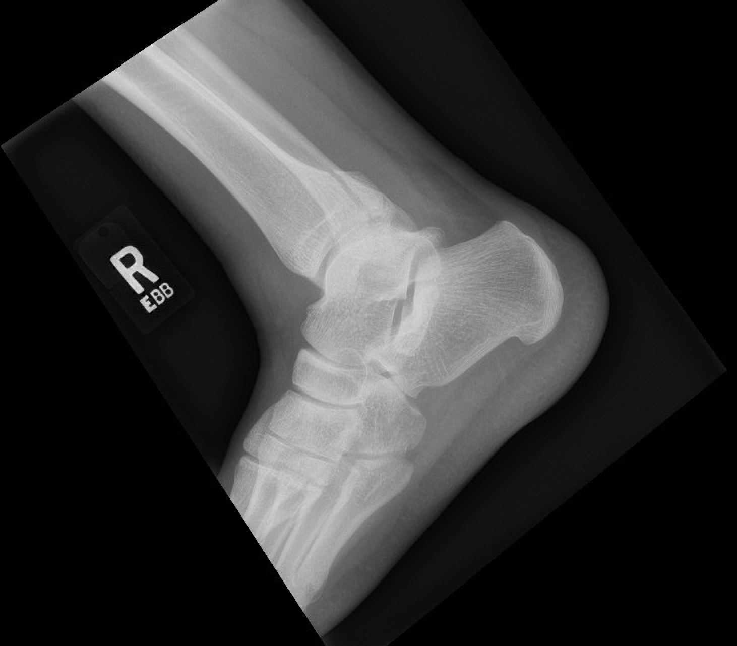
 A 32-year-old female presents to the emergency department with right foot pain after rolling her ankle during dance class. On exam, there is mild swelling and tenderness over the 5th metatarsal. The above x-ray was obtained (Figure 1: Case courtesy of Dr. Henry Knipe, Radiopaedia.org, rID:
 A 67-year-old male with a history of bilateral total hip arthroplasties (THA) several years ago presents with left hip pain after a fall. He was walking downstairs and slipped, twisting his leg internally and with adduction and flexion of the hip to catch himself. He denies falling but felt an immediate pop in his left hip and could no longer bear weight. AP and lateral radiographs of the pelvis and left hip were obtained and are shown above (Image 1. Case courtesy of Dr Andrew Taylor, Radiopaedia.org, rID: