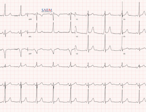
 Posterior myocardial infarction (MI) represents 3.3 – 21% of all acute MIs and can be difficult to diagnose by the standard precordial leads. Typically, leads V7 – V9 are needed to diagnose this entity. Luckily, leads V1 – V3, directly face the posterior wall of the left ventricle and are the “mirror image” of the posterior wall of the left ventricle.
What is the significance of ST depression in leads V1 – V3?
This suggests that there is evidence of:
- Anterior wall ischemia, or
- Posterior wall MI
What are ECG criteria for posterior MI on the standard 12-lead ECG? 1
- R/S wave ratio >1.0 in lead V2
- Co-existing acute, inferior, and/or lateral MI
- Limited to leads V1 – V3:
- ST-segment depression (horizontal moreso than downsloping or upsloping)
- Prominent R wave
- Prominent, upright T wave
- Combination of horizontal ST-segment depression with upright T wave
What is the correct placement of leads V7 – V9? 2
- V7: posterior axillary line
- V8: inferior angle of the scapula
- V9: just to the lateral to the vertebrae

How is the “Flip Test” Performed?
- Get a standard 12 lead ECG
- Turn it over 180 degrees to look at the back of the upside-down paper.
- Aim the paper at a bright light source to enable seeing the “flipped” tracings.
- ST elevation in these leads V1 – V3 with Q waves is consistent with posterior STEMI

Standard 12-lead ECG with abnormal findings circled in red

Same flipped 12-lead ECG with same V1-V3 abnormality highlighted in red circles.
Posterior MI: Anterior R waves versus Posterior Q waves on ECG
With a posterior MI, R waves in the anterior leads (V1 – V3) and Q waves in the posterior leads (V7 – V9) can be present, but how good is the correlation between the two? In a case series of 58 patients with posterior MIs 3 :
- 44.4% of patients had anterior lead R waves
- 44.4% of patients had posterior lead Q waves
- Only 50% of patients with posterior MI had both
Conclusion: There is poor correlation between anterior R waves and posterior Q waves.
Posterior MI: Anterior ST-depression versus Posterior ST elevation on ECG
Again with posterior MI, ST-depressions in the anterior leads (V1 – V3) and ST-elevations in the posterior leads (V7 – V9) can be present, but how good is the correlation between the two? In case series of posterior MIs:
- 61 – 91.67% had ST depression in anterior leads 3,4
- 91 – 100% had ST elevation in posterior leads 3,4
- 84.6% of patients with posterior MI had both anterior ST-depressions and posterior ST-elevations 3
Conclusion
A cheap and easy way to diagnose a posterior MI is flipping the ECG over and looking at leads V1 – V3 in the light, but using posterior leads (V7 – V9) will more accurately diagnose patients with posterior MI.
I would like to thank Dr. Gemma Morabito (@MedEmIt) for the idea of this post and Amal Mattu (@amalmattu) for these ECGs.


