

An otherwise healthy 34-year-old male presented to the Emergency Department with two weeks of anterior neck pain. Symptoms began with several days of pain in his mandibular molars, progressing to pain and swelling in the neck. In the last several days, the patient developed warmth and redness in the chest wall associated with subjective fever and chills. Additionally, the patient reports difficulty swallowing solid foods secondary to odynophagia associated with intermittent globus sensation. He has no history of immunocompromise and denies any drug or alcohol use. Of note, he has not seen a dentist in many years.

Vitals: BP 115/80; HR 120; T 101°F; RR 16; O2 sat 97%
General: Well appearing in no acute distress
HEENT: Poor dentition, mild trismus. No gingival inflammation or swelling or induration to suggest abscess. The floor of the mouth is unremarkable.
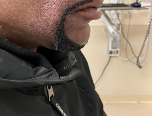
Skin: The neck and upper chest demonstrate erythema and tenderness with an enlarged area of fluctuance on the superior aspect of the left breast (Figure 1).
White blood cell (WBC) count: 6.3 k/uL
Lactate: 1.6 mmol/L
Glucose: 95 mg/dL
Creatinine: 0.72 mg/dL
Lemierre Syndrome, also known as septic thrombophlebitis of the internal jugular vein, is a rare condition with an incidence of 3-15 cases per million people. This condition occurs when an oropharyngeal or odontogenic infection spreads locally from pharyngeal tissue to the internal jugular vein. The pathogens classically arise from normal oral flora, most commonly Fusobacterium necrophorum. The presentation may be associated with trismus and/or dysphagia. Subsequent complications, including localized abscess formation and bacteremia, stem from a combination of surrounding tissue invasion and systemic septic embolization.
Given the potential for regional lymphatic spread and septic embolization, patients may present with both local and systemic findings. Skin exam may reveal regionalized cellulitic or infectious changes overlying the neck or chest (Figure 1).
Respiratory signs and symptoms may suggest the presence of pulmonary septic emboli or mediastinitis.
Constitutional symptoms including fever, chills, and fatigue are common though nonspecific. The differential is broad and includes a number of infectious, lymphatic, endocrine, and neoplastic conditions.
It is essential for the clinician to consider the alternative diagnosis of Ludwig’s Angina through careful evaluation of the oral floor.
Given the potential for oropharyngeal and respiratory compromise, emergency clinicians must maintain a high index of suspicion for this condition. Diagnostics should include laboratory studies with blood cultures, as well as CT imaging of the neck and chest to evaluate for filling defects of the internal jugular vein.
When entertaining the diagnosis, early antibiosis is prudent. Treatment should include both an extended course of antibiotic therapy as well as surgical source control of abscesses. Given the propensity for thrombus development (Figure 2), anticoagulation may be considered, but its indication here remains controversial. Patients with Lemierre Syndrome will require surgical consultation and hospital admission.
Take-Home Points
- Lemierre Syndrome is a septic thrombophlebitis of the internal jugular vein most commonly occurring via direct spread from the oral cavity. Distinction from Ludwig’s Angina is imperative.
- Given the proximity to critical structures and the potential for systemic organ dysfunction from septic emboli, emergency physicians need to maintain a high clinical suspicion for this rare diagnosis.
- Treatment includes parenteral antibiotics and prompt consultation of medical and surgical subspecialists to identify the infectious source as well as mitigate against systemic spread and/or thrombus propagation.
- Kuppalli K, Livorsi D, Talati NJ, Osborn M. Lemierre’s syndrome due to Fusobacterium necrophorum. Lancet Infect Dis. 2012 Oct;12(10):808-15. doi: 10.1016/S1473-3099(12)70089-0. Epub 2012 May 25. PMID: 22633566.

Andrew Mittelman, MD
Latest posts by Andrew Mittelman, MD (see all)
- SAEM Clinical Images Series: A Grain of Sand… or Something More Sinister? - February 27, 2026
- SAEM Clinical Images Series: A Pedunculated Bone to Pick - January 12, 2026
- SAEM Clinical Images Series: Tangled in the Toilet - October 31, 2025

Laura Welsh, MD
Department of Emergency Medicine
Boston Medical Center
Latest posts by Laura Welsh, MD (see all)
- SAEM Clinical Images Series: A Serious Pain in the Neck - October 16, 2023
- SAEM Clinical Images Series: A Painful Swollen Digit - August 21, 2023

Felisha Gonzalez, MD
Boston Medical Center
Latest posts by Felisha Gonzalez, MD (see all)
- SAEM Clinical Images Series: A Serious Pain in the Neck - October 16, 2023

Jordan Spector, MD
Residency Program Director
Department of Emergency Medicine
Boston Medical Center
Latest posts by Jordan Spector, MD (see all)
- SAEM Clinical Images Series: Ptosis? A Don’t Miss Diagnosis! - April 4, 2025
- SAEM Clinical Images Series: A Serious Pain in the Neck - October 16, 2023
- SAEM Clinical Images Series: A Painful Swollen Digit - August 21, 2023

