

The patient is a 60-year-old male with a history of insulin-dependent diabetes, hypertension, and hyperlipidemia who presents to the Emergency Department after one day of sudden onset right eye pain associated with nausea and vomiting. He notes progressively blurring vision and vision loss in his right eye since the onset of the pain. His wife noted redness of his sclera and urged him to go the emergency department. He can now only sense light and shadows with his right eye. He denies traumatic injury or any history of serious ophthalmological pathology. He wears corrective eyeglasses and does not use contacts. He has no other complaints at this time.

Vitals: BP 149/83; HR 107; R 17; T 98.9°F; O2 sat 100 on room air.
General: Appears to be in pain and uncomfortable.
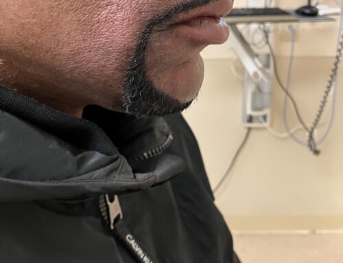
HEENT: As shown. Extraocular movements are intact. The right pupil is fixed and dilated with a relative afferent pupillary defect. There is no sign of traumatic injury.
Neck: There are no carotid bruits auscultated.
Cardiovascular: Regular rate and rhythm, no murmur.
Neurologic: Normal other than the abnormal findings of the right eye.
Imaging: POCUS of the right eye is performed, image as shown.
Acute angle-closure glaucoma.
Ultrasound shows retinal detachment with subretinal hemorrhage and associated choroidal detachment.
Acute angle-closure glaucoma occurs due to a rapid increase in intraocular pressure (IOP) due to outflow obstruction of the aqueous humor. Patients with a shallower angle between the iris and the cornea in the anterior chamber are predisposed to this condition. This is characterized clinically by severe eye pain, headache, nausea, vomiting, blurred vision, and multicolored halos around lights. If left untreated, this can result in optic neuropathy and vision loss. The diagnosis of acute angle-closure glaucoma is confirmed with elevated intraocular pressure (IOP) measurements obtained via tonometry. Normal IOPs are between 10 and 21 mmHg. The pressure in this patient’s right eye was 47 mmHg. Slit-lamp microscope exam showed a shallow anterior chamber, corneal edema, fixed dilated pupil, and conjunctival injection around the limbus (ciliary flush). Uncommonly, retinal and choroidal detachment may cause secondary acute angle-closure glaucoma, as seen in this case. Treatment includes medical and surgical interventions to reduce IOP, address underlying causes, and manage associated pain and nausea.
Take-Home Points
Retinal detachments are seen as a “V”-shaped hyperechoic and freely moving membrane tethered to the optic disc on ultrasound.
Acute angle-closure glaucoma is an ocular emergency. Delays in treatment can result in optic neuropathy and permanent vision loss.
- Stenberg RT, Nelson J, Rabinowitz J, Simon EL. Spontaneous Hyphema and Vitreous Hemorrhage Causing Secondary Glaucoma in a Patient on Apixaban. J Emerg Med. 2023;64(3):359-362. doi:10.1016/j.jemermed.2022.12.021
- Jersey A, Perice L, Li N, Johnson J, Dulani T. Acute Angle-Closure Glaucoma Secondary to Vitreous Hemorrhage Diagnosed with the Aid of Point-of-Care Ultrasound. J Emerg Med. 2020 Dec;59(6):e235-e237. doi: 10.1016/j.jemermed.2020.08.015. Epub 2020 Sep 29. PMID: 33004244.
- Chen SN, Ho CL, Ho JD, Guo YH, Chen TL, Chen PF. Acute angle-closure glaucoma resulting from spontaneous hemorrhagic retinal detachment in age-related macular degeneration: case reports and literature review. Jpn J Ophthalmol. 2001 May-Jun;45(3):270-5. doi: 10.1016/s0021-5155(00)00382-8. PMID: 11369377.

Milap Desai, MD
York WellSpan Hospital
Latest posts by Milap Desai, MD (see all)
- SAEM Clinical Images Series: Painful Red Eye - March 20, 2026
