

A 55-year-old female presented with the complaint of “my right eye popped out.” Symptoms started approximately seven hours prior to arrival and progressive, severe pain eventually prompted her visit to the ED. This happened once 10 years ago, requiring reduction in the ED. The patient denied preceding trauma, rubbing her eyes/eye-lids, or any history of thyroid disease. She endorsed right eye blurred vision and severe pain.

Vitals: HR 86; RR 16; SpO2 97% on room air; BP 179/111
General: Appears uncomfortable
Head: Atraumatic
Ocular:
OD: globe luxation with severe injection and chemosis. Severe corneal dryness. Pupil appears 3mm, minimally reactive, though poor view of pupil secondary to exposure keratopathy. Upper and lower lids inverted beneath globe. IOP 21. Unable to assess extraocular movements
OS: appears grossly normal. Pupil 3mm and reactive. Full extraocular movements.
Thyroid stimulating hormone and thyroxine within normal limits.
Spontaneous globe luxation (SGL). Luxation of the globe is characterized by the anterior displacement of the globe beyond the orbital rim [1], as seen in photo one. Though SGL is a rare condition, risk factors include proptosis, shallow orbits, or space-invading retrobulbar lesions [2]. Case study reports have also indicated trauma and frequent eyelid manipulation as causes of globe luxation [1].
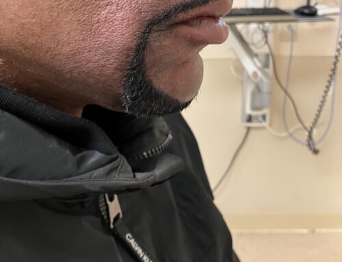
After assuring adequate anxiolysis and analgesia with IV medications and tetracaine eye drops, the patient should be placed in a supine position. To reduce the globe, the eyelids should be extracted from behind the globe and retracted outwards while direct and even pressure is applied to the globe with damp gauze. For our patient, a lateral tarsorrhaphy was performed by ophthalmology at the bedside given severe keratopahy, lagopthalmos (as seen in photo two), and re-subluxation with Valsalva. Given the unknown etiology of the luxation, thyroid laboratory testing and orbit computed tomography were performed, which were unremarkable. The patient was discharged from the emergency department with tobradex ointment and ophthalmology follow-up in one week
Take-Home Points
-
Immediate reduction of a luxed globe is paramount.
-
Consider topical anesthetic drops and IV analgesia and/or anxiolytics to help assist with patient discomfort and dry eye.
-
Consider labs and imaging to assess for any underlying etiology of spontaneous globe luxation.
-
Kelly, E.W. and Fitch, M.T. (2013) Recurrent Spontaneous Globe Subluxation: A Case Report and Review of Manual Reduction Techniques. Available at: https://www.sciencedirect.com/science/article/abs/pii/S0736467911011462 (Accessed: 04 January 2024).
-
Yadete, T. et al. (2021) Spontaneous globe subluxation: A case report and review of the literature – international journal of emergency medicine, BioMed Central. Available at: https://intjem.biomedcentral.com/articles/10.1186/s12245-021-00398-x (Accessed: 04 January 2024).

Rachel Whittaker, MD
University of Chicago
Latest posts by Rachel Whittaker, MD (see all)
- SAEM Clinical Images Series: Spontaneous Eye Luxation - January 13, 2025

Emily Jameyfield, MD
University of Chicago
Latest posts by Emily Jameyfield, MD (see all)
- SAEM Clinical Images Series: Spontaneous Eye Luxation - January 13, 2025
