

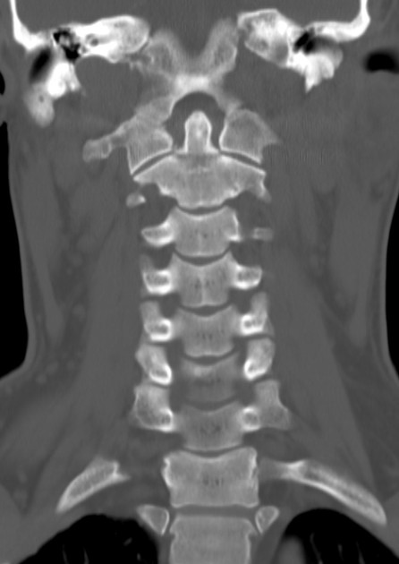
80-year-old male patient brought in by ambulance for a witnessed fall. A cervical collar was placed by EMS because of midline neck pain. The patient is neurologically intact. A CT of the cervical spine was obtained and is shown above (Case courtesy of Dr. Talal F M Abdullah, Radiopaedia.org, rID: 58030).
Odontoid fracture, type II. The odontoid process, or dens, is a superior bony projection of C2 that, along with the transverse ligament, is a key stabilizer of C1.

Figure 2. Case courtesy of Dr. Talal F M Abdullah, Radiopaedia.org, rID: 58030. Annotations by author, fracture highlighted in red
- Pearl: Odontoid fractures can occur in elderly patients via a low energy mechanism like ground-level falls. They may also occur in younger patients during high-energy trauma that causes hyperflexion or hyperextension.
Type I: Alar ligament avulsion fracture of the apex. Stable
Type II: Fracture at the waist of the odontoid process. Unstable
Type III: Fracture of the body of C2 with involvement of the lateral masses. Unstable

Figure 3. Case courtesy of Dr. Mohammad Taghi Niknejad, Radiopaedia.org, rID: 21310. Odontoid fractures type 1, 2, and 3 from left to right
- Pearl: Type II fractures have a high nonunion rate because the waist of the odontoid process has limited blood supply for two reasons [1]:
- Vascular supply arrives from the apex and the base, forming a watershed area at the waist.
- The waist lacks cancellous bone, which contains a highly vascularized meshwork of bone and blood vessels that allows for faster healing.
The patient should be placed in a cervical collar and maintained in strict spinal precautions. A spine surgeon should be consulted emergently as guided by institutional protocols. MRI can be obtained if neurologic symptoms are present.
- PEARL: Management typically includes the following
- Type I -> hard cervical collar for 6-12wk.
- Type II -> operative management (screw fixation or C1-2 posterior fusion) [1-2].
- Type III -> hard cervical collar or halo immobilization [1].
Resources & References:
Check out this Paucis Verbis card to review cervical spine imaging rules.
- Egol K, Koval, KJ, Zuckerman JD. Handbook of Fractures. Lippincott Williams & Wilkins. 2010 ISBN: 160547760.
- Gembruch O, Lemonas E, Ahmadipour Y, Sure U, El Hindy N, Dodel R, Müller O. Treatment of Odontoid Type II Fractures in Octogenarians: Balancing Two Different Treatment Strategies. Neurospine. 2019 Jun;16(2):360-367. Epub 2019 Feb 23. PMID: 31154696.

BuMin Kong, MD
Department of Emergency Medicine
Loma Linda University Medical Center
Latest posts by BuMin Kong, MD (see all)
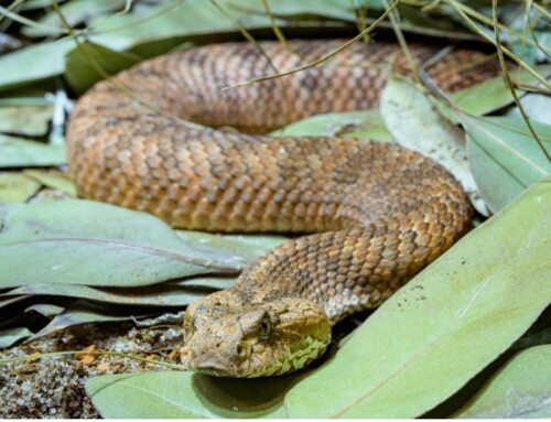
- SplintER Series: Attack by Bat - June 16, 2021
- SplintER Series: The Tooth of the Cervical Spine - June 9, 2021
- SplintER Series: Diver’s Nightmare - May 12, 2021

Mark Hopkins, MD
Latest posts by Mark Hopkins, MD (see all)
- SplintER Series: Patellar Tendon Rupture - January 25, 2023
- SplintER Series: Don’t forget about the (tibial) spine! - December 28, 2022
- SplintER: Pop, Lock & Drop It - November 9, 2022

Alexander J. Tomesch, MD
Department of Orthopedic and Sports Medicine
University of Arizona - Tucson
Latest posts by Alexander J. Tomesch, MD (see all)
- SplintER Series: En Pointe - January 11, 2023
- SplintER Series: Don’t forget about the (tibial) spine! - December 28, 2022
- SplintER Series: Hip, Hip, Hooray! - December 16, 2022
