
 Two patients present to your emergency department: Patient 1 is a 17 year-old soccer player who fell during a game onto their right side and is now complaining of mild right shoulder pain. You obtain x-rays (Figure 1). Patient 2 is a 21 year-old motorist who lost control and went over the handlebars. They heard a pop and are complaining of left shoulder pain. You obtain shoulder x-rays (Figure 2). For these cases, what are your diagnoses, expected physical examination findings, and emergency department management?

Figure 1: Image courtesy of Dr Benoudina Samir, Radiopaedia.org

Figure 2: Image courtesy of Dr Craig Hacking, Radiopaedia.org
- Acromioclavicular (AC) Joint Separation:
- Case 1 (Figure 1): Type II AC joint separation
- Case 2 (Figure 2): Type IV AC joint separation.
- Pearl: Because this dislocation is primarily posterior, it can be easily missed on an AP film. An axillary view (Figure 3) is typically required to make the diagnosis.

Figure 3: Axillary view showing clavicle (dashed line) displaced posteriorly in comparison to the acromion (solid line) in a Type IV. Image courtesy of Dr Bijoor, Radiopaedia.org, edits by Dr Luz Silverio
- AC joint separations can occur from blunt trauma to the shoulder, most commonly from falling onto a shoulder with an adducted arm [1]. This forces the acromion inferomedially and the clavicle superiorly.
- AC joint separations are classified using the Rockwood Classification System [2]. It grades the degree of separation of both the AC ligament and the coracoclavicular (CC) ligament. See Table 1 and Figure 4.
| Type | Description |
|---|---|
| I | AC sprain, normal radiograph |
| II | AC torn, CC sprained, clavicle does not elevate above the superior border of the acromion |
| III | AC and CC torn; clavicle is above the superior border of the acromion and <25mm of displacement measured at the CC space. |
| IV | AC and CC torn; posterior displacement of the clavicle. |
| V | AC and CC torn; >25mm of superior displacement of the clavicle at the CC space. |
| VI | AC and CC torn; distal clavicle is displaced inferiorly and underneath the coracobrachialis/biceps tendon. Very rare. |
Table 1: Rockwood Classification for AC joint separation

Figure 4: Normal Acromioclavicular (AC) and Coracoclavicular (CC) Spaces. AC Space should be 5-8 mm (red). CC Space (green) should be 10-13 mm. Image courtesy of Dr Jeffrey Hocking, Radiopaedia.org
- A patient with an AC dislocation will have tenderness at the distal clavicle and AC joint and an abnormal contour of the affected shoulder when compared contralaterally. There may be referred pain to the trapezius.
- Pearl: Consider other etiologies that may mimic an AC separation – clavicle fracture, shoulder dislocation, rotator cuff tear, pneumothorax, superior labral antero-posterior (SLAP) lesion of the shoulder, or cervical radiculopathy.
- Plain films of the shoulder and clavicle. Ensure an axillary view is performed to evaluate for a type IV.
- Pearl: Contralateral films may be helpful to improve classification [3].
- Type I to II are managed conservatively with rest, ice, and analgesics.
- Pearl: Sling and encourage early mobilization as tolerated to avoid adhesive capsulitis.
- Type III injuries may require operative intervention for overhead laborers, elite athletes, or patients with greater than 20mm of CC space.
- Types IV to VI injuries are managed operatively with AC joint fixation or ligament reconstruction [4, 5].
- Case 1 (Type II AC): Type I to III AC separations can be discharged with close sports medicine/orthopedic follow-up. Encourage early range of motion. Counsel that it can take approximately 6 weeks to regain functional motion and approximately 12 weeks to regain normal activity. Physical therapy may be required.
- Case 2 (type IV AC): Type IV to VI AC joint injuries require operative repair. Orthopedic consultation is recommended to facilitate operative planning.
For more information about acromioclavicular separations, University of Washington has a great AC joint reference and Orthobullet’s review of acromioclavicular separations is also worth checking out. Want more SplintER?
For more cases like these, you can subscribe to the Ortho EM Pearls email series hosted by Drs. Will Denq, Tabitha Ford, and Megan French, who have kindly shared some of their content with ALiEM.
References:
- Vanhoenacker F, Maas M, Gielen JL. Imaging of Orthopedic Sports Injuries. Springer Verlag. (2006) ISBN:3540260145
- Kiel J, Kaiser K. Acromioclavicular Joint Injury. In: StatPearls. Treasure Island (FL): StatPearls Publishing; 2019 jan. PMID: 29630240
- Ibrahim ef, Forrest NP, Forester A. Bilateral weighted radiographs are required for accurate classification of acromioclavicular separation: An observational study of 59 cases. Injury Int J Care Injured 46(2015)1900-1905. PMID: 26194267
- Stapczynski, J S, and Judith E. Tintinalli. Tintinalli’s Emergency Medicine: A Comprehensive Study Guide, 8th Edition. New York: McGraw-Hill Education, 2016. ISBN: 9780071794763
- Favorito, PJ., Herbst., KA., Acromioclavicular joint injuries, Shoulder and Elbow Trauma and its Complications., Woodhead Publishing Series in Biomaterials., Volume 1., Pages 215-231., 2015. ISBN: 9781782424727

Sergio Alvarez, MD CAQSM
Department of Orthopedics
Sutter Health/Palo Alto Medical Group
Latest posts by Sergio Alvarez, MD CAQSM (see all)
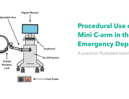
- Procedural Use of a Mini C-arm in the Emergency Department - December 31, 2025
- SplintER Series: Two cases of shoulder pain - February 5, 2020
- SplintER Series: Funny Looking Finger - August 30, 2019

Megan French, MD, FACEP
Utah Emergency Physicians
Latest posts by Megan French, MD, FACEP (see all)
- SplintER Series: Two cases of shoulder pain - February 5, 2020
- SplintER Series: The Recurrent Shoulder Dislocation - January 22, 2020
- SplintER Series: A Rare Cause of Traumatic Thumb Pain - December 30, 2019

William Denq, MD CAQ-SM
Department of Emergency Medicine
University of Arizona
@willdenq
Latest posts by William Denq, MD CAQ-SM (see all)
- SplintER Series: Fracture After a Fall From a Bunk Bed - August 6, 2021
- SplintER Series: Open Fracture - May 28, 2021
- SplintER Series: What is Wrong With My Daughter? - May 3, 2021