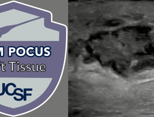
Welcome to another ultrasound-based case, part of the “Ultrasound For The Win!” (#US4TW) Case Series. In this case series, we focus on a real clinical case where bedside ultrasound changed the management or aided in the diagnosis. In this case, a 63-year-old man presents with a painful, warm, and erythematous area of his abdomen.
Case Presentation
A 63-year-old man with history of diabetes, hypertension, and hyperlipidemia presents with a painful area on his right lower abdomen. He states he noticed pain and redness today, and that it has been worsening over the course of the day. He denies any previous history of similar symptoms and denies trauma. On physical examination, he is a morbidly obese gentleman, in no acute distress. Examination of his abdomen reveals a 10 cm x 12 cm erythematous and tender area on the surface of the right side of his lower abdomen. The area is warm to touch without fluctuance or crepitus. Genitourinary examination is unremarkable.
Vitals
BP 173/82 mmHg
P 111 bpm
RR 23 breaths/min
O2 97% room air
T 37.9 C
Differential Diagnosis
- Abscess
- Cellulitis
- Necrotizing Fasciitis
Laboratory Investigations
- Total White Blood Cell count: 18 x mm3
- C-Reactive Protein: 240 mg/L
- Hemoglobin: 14.3 g/dL
- Sodium: 139 mmol/L
- Creatinine: 119 umol/L (or 1.35 mg/dL)
- Glucose: 12 mmol/L (or 216 mg/dL)
- Lactate: 4.1 mmol/L
Point-of-care Ultrasound

Figure 1. Cobblestoning of subcutaneous soft tissue with fluid in the deeper fascial plane

Figure 2. Another view of the cobblestoning of subcutaneous soft tissue and fluid in the deep fascial plane.

Figure 3. Cobblestoning of the subcutaneous tissue (#) and fluid in the deep fascial plane (arrow) is seen.
Ultrasound Image Quality Assurance
The ultrasound images were obtained using the high-frequency linear probe, which is beneficial when attempting to visualize superficial structures within a few centimeters from the surface. The images reveal cobblestoning of the subcutaneous tissue, a non-specific finding that can be seen with cellulitis (Figure 1, Figure 2). Of note, the subcutaneous tissue is uniformly thickened; a comparison of a normal area (e.g. a contralateral limb) can be visualized to confirm abnormal thickening. Deep to the subcutaneous layer is the deep fascial plane, where abnormal fluid is seen in this case [Fig. 3]. These findings can be seen with necrotizing fasciitis. As the disease progresses, abnormal air, visualized as “dirty shadowing” on ultrasound, may be seen in late and more severe cases.
Disposition and Case Conclusion
Given the concerning history and physical examination along with the point-of-care ultrasound concerning for necrotizing fasciitis, empiric antibiotics (IV vancomycin and piperacillin/tazobactam) were given, and surgery was consulted.
The patient was taken to the operating room where a wash out and debridement was performed with a confirmed diagnosis of necrotizing fasciitis. The patient was monitored in the intensive care unit post-operatively and has since been discharged and is doing well.
Background on Necrotizing Fasciitis
Necrotizing fasciitis is rare (with an incidence of 4.3 infections per 100,000 in the United States), but severe soft tissue infection1,2. The most severe form of soft tissue infections, necrotizing fasciitis is a rapidly progressing infection of the subcutaneous tissue and fascia that is potentially limb and life threatening, with a mortality rate of up to 76%2,3. Bacterial enzymes cause tissue necrosis, leading to fluid that can be visualized in the deep fascial layer. The typical bacterial pathogens involved in necrotizing fasciitis include staphylococci, streptococci, and anaerobes, and antibiotic coverage should provide broad coverage for these organisms4. Definitive management requires operative debridement and potential fasciotomy.
The classic physical examination findings of necrotizing fasciitis, including a rapidly progressing area of erythema with ill-defined borders, are often indistinguishable from other soft tissue infections including cellulitis and abscess, especially early in the disease process. Thus, a high index of clinical suspicion is required in the Emergency Department2. While physical exam findings including blistering, hemorrhagic bullae, and crepitus can increase the suspicion of necrotizing fasciitis, these are often late findings seen only in severe and progressed cases2. While necrotizing fasciitis is considered a clinical diagnosis, there may be some utility for laboratory tests and point-of-care ultrasound to aid in risk-stratifying equivocal cases.
LRINEC Score
The LRINEC (Laboratory Risk Indicator for Necrotizing Fasciitis) score utilizes 6 common laboratory tests to risk stratify patients with concern for possible necrotizing fasciitis (Table 1)3. A score of ≥6 should raise your suspicion of the diagnosis, while a score of ≥8 is strongly predictive of necrotizing fasciitis3. In this case, the LRINEC score is 6, which increases the suspicion of necrotizing fasciitis.
Table 1. Laboratory Risk Indicator for Necrotizing Fasciitis (LRINEC) Score
| LRINEC Score > 6 should raise suspicion of necrotizing fasciitis. Score > 8 is strongly predictive of necrotizing fasciitis. (Modified from Wong et al.) | |
| Lab, Units | Score |
| C-Reactive Protein, mg/L | |
| < 150 | 0 |
| ≥ 150 | 4 |
| White cell count, per mm3 | |
| < 15 | 0 |
| 15 – 25 | 1 |
| > 25 | 2 |
| Hemoglobin, g/dL | |
| > 13.5 | 0 |
| 11 – 13.5 | 1 |
| < 11 | 2 |
| Sodium, mmol/L | |
| ≥ 135 | 0 |
| < 135 | 2 |
| Creatinine | |
| ≤ 141 mmol/L or 1.6 mg/dl | 0 |
| > 141 mmol/L or 1.6 mg/dL | 2 |
| Glucose | |
| ≤ 10 mmol/L or 180 mg/dL | 0 |
| > 10 mmol/L or 180 mg/dL | 1 |
Ultrasound Findings for Necrotizing Fasciitis
Ultrasound can also be used to identify patients with necrotizing fasciitis. While CT and MRI have been the more traditionally used imaging modalities, they are time consuming, costly, and delay the time to definitive operative management. The ultrasonographic findings of necrotizing fasciitis include diffuse thickening of the subcutaneous tissue when compared to the contralateral side or limb, and a layer of fluid seen more than 4 mm deep along the deep fascial layer1. Using these criteria, ultrasound has been shown to be 88.2% sensitive, 93.3% specific, and 91.9% accurate1. As the disease progresses, air within the fascial layer, seen as “dirty shadowing” may be seen. A useful mnemonic has been described in the literature as the STAFF exam (Subcutaneous Thickening, Air, and Fascial Fluid)2.
Necrotizing fasciitis remains a clinical diagnosis, and concern for the disease requires prompt surgical consultation. While laboratory tests (LRINEC score) and ultrasound are beneficial and can aid in the risk stratification and diagnosis of cases, they should not be used solely to rule out the disease.
Take Home Points
- Necrotizing fasciitis is a rare but potentially limb and life threatening infection, requiring a high index of clinical suspicion.
- While necrotizing fasciitis is a clinical diagnosis, the LRINEC score and point-of-care ultrasound can aid in the risk-stratification and early diagnosis of the disease.
- Ultrasonographic findings suggestive of necrotizing fasciitis include:
- Fascial and subcutaneous thickening
- Fluid in the deep fascial layer
- Subcutaneous air
*Note: All identifying information and certain aspects of the case have been changed to maintain patient confidentiality and protected health information (PHI).


