Separation of the acromioclavicular (AC) joint is a common injury encountered in the emergency department. Keys to optimal outcome and return of function in these patients include knowledge of injury mechanism, diagnosis and classification, and initial treatment.
Case
A 16-year-old male presents complaining of right shoulder pain. The patient is the quarterback for his high school football team. He was tackled, falling onto his right shoulder. He appears uncomfortable, but his vital signs are within normal limits. A focused exam of the right shoulder reveals limited range of motion due to pain and tenderness over the distal clavicle and acromion. There is no deformity or palpable fracture, however, the clavicle is slightly mobile in an anterior direction. An x-ray is obtained due to your concern for a possible AC joint separation.
What is the AC joint?
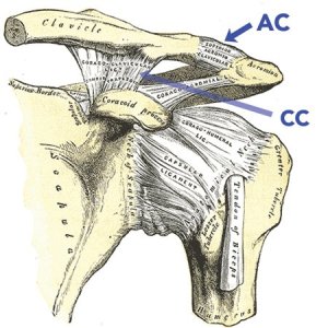
The acromioclavicular joint is a synovial articulation between the distal clavicle and the acromion. Like other joints, stability of this joint is provided by a capsule and ligaments. There are the acromioclavicular (AC) ligaments and the coracoclavicular (CC) ligaments. The AC ligaments resist anterior-posterior movement, while the CC ligaments resist movement in the superior-inferior direction. On clinical exam, directional instability of the clavicle occurs with varying degrees of ligamentous injury. It is also thought that the deltoid and trapezius muscles provide dynamic stability for this joint.
Anterior view of L shoulder
How do you diagnose an AC joint separation?
Mechanism: This is generally caused by a force through the shoulder, driving the acromion inferiorly. This can occur during contact sports, falls onto the shoulder or arm, or with multidirectional forces.
Physical Exam: The patient will have localized pain and possibly a deformity. Compression of the joint and horizontal adduction of the arm will elicit joint mobility. The joint may also be unstable without these tests.
Imaging: This aids in the diagnosis. Plain films are the most common modality, but bone scan, ultrasound, and magnetic resonance imaging may also be used. Stress films are often mentioned in the literature, but are not always worthwhile to obtain in the emergency department.
Classification of AC joint separations
This is made based on the Rockford grades:
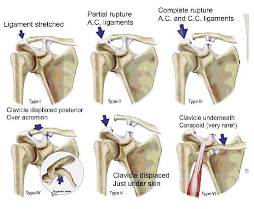
- Grade I: Sprain of the AC ligaments, but the ligaments are intact. There is no instability of the clavicle on exam and the patient will have a normal x-ray.
- Grade II: Involves rupture of the AC ligaments, but the CC ligaments remain intact. The clavicle will have some mobility in the anterior-posterior direction. On x-ray, the lateral clavicle may be slightly elevated. Stress films, however, will show normal alignment of the AC joint.
- Grade III: Complete disruption of the AC and CC ligaments without significant disruption of the delto-trapezial fascia. A deformity is present on exam with the clavicle appearing elevated and unstable in both vertical and horizontal directions. This separation of the clavicle and acromion is seen on x-ray.
- Grade IV: The distal clavicle is posteriorly displaced into the trapezius muscle. A posterior deformity is present on exam. On plain films, the clavicle will be displaced posteriorly on axillary views.
- Grade V: A more severe form of grade III. Not only is there disruption of the AC and CC ligaments, but there is also disruption of the delto-trapezial fascia. There may be tenting or pseudo elevation of the lateral clavicle, due to downward displacement of the scapula. On x-ray, there will be a 2-3 times increase in the distance between the coracoid and clavicle or a 100-300% increase in the clavicle-acromion distance.
- Grade VI: Involves inferior displacement of the distal clavicle, either subacromial or subcoracoid. This is a result of severe trauma and there are often other significant injuries present.
How do you treat AC joint separations?
First, it should be noted that there are no randomized controlled trials on the management of AC joint separations. It is generally accepted that the objectives of management should be to reduce the risk of complications following injury, restore function, and optimize sporting performance. The consensus in the literature is that Grade I-III injuries can be managed conservatively, while Grade IV-VI injuries need surgical intervention, usually urgently, if not emergently. However, there was one study by Lizaur et al that had 46 patients with Grade III injuries. 1 Of those patients, 94% had a concurrent injury to the delto-trapezial muscle complex, needing surgical repair. This has sparked some debate in the realm of orthopedics.
Acute phase of AC joint separations: Place the injured extremity in a sling and providing analgesia and anti-inflammatory modalities. These patients will need referral to an orthopedic surgeon for further management, including some form of a rehab program. The best practice for an optimal outcome in these patients involves providers with a working knowledge of anatomy, kinematics, kinetic chain concepts, the healing continuum, exercise physiology, and the ability to adapt treatment strategies. Gladstone et al published the most common protocol for treatment and rehabilitation in 1997, which involves progressive range of motion and strengthening exercises along with assessment for return to activity. 2
In the emergency department, we can provide the initial immobilization and pain control. It is probably just as important to provide a realistic timeframe for recovery to these patients. Time based guidelines for return to sport or activity are designed to indicate when recovery is falling out of the ideal time. Recommendations are 3 :
- 2-4 weeks for Grade I injuries
- 4-6 weeks for Grade II injuries
- 6-12 weeks for Grade III injuries
Case conclusion
Due to normal appearing x-ray, you diagnose the patient with a Grade II AC joint separation (due to the clavicle mobility on exam). The patient is provided analgesia and placed in a sling for comfort. You inform the patient it will likely take at least 4 weeks for full recovery, and arrange orthopedic follow up.
Bottom line
AC joint separations are common in the ED. Use a focused physical exam and imaging to make the diagnosis. Generally, Grade I-III injuries can be managed conservatively with early range of motion exercises and orthopedic follow up. Grade IV-VI injuries need an orthopedic consultation in the emergency department. Place patients in a sling and provide pain control.

