

The patient is a 40-year-old female who presents to the Emergency Department with bilateral leg swelling. Her symptoms started six days prior and have progressively worsened. Her symptoms are associated with shortness of breath with no chest pain. The patient has taken an over-the-counter diuretic, which has helped with her symptoms. She also reports intermittent vaginal bleeding for the past two months, with a LMP that was two months prior. She is not currently on contraceptives, and does endorse unprotected intercourse over this time. The patient denies headache, blurry vision, nausea or vomiting, abdominal pain, urinary complaints, diarrhea or constipation. She has no other complaints at this time.


Vitals: BP 140/86; HR 97; R 14; T 99°F; O2 sat 99% on room air.
General: Well appearing, no acute distress.
Respiratory: Clear to auscultation.
Cardiovascular: Regular rate and rhythm, no murmur.
Abdomen: Soft, nondistended, nontender.
Extremities: Trace bilateral pitting edema. Normal range of motion, neurovascularly intact, equal pulses bilaterally.
Neurological: No focal neurological deficits.
Hgb: 9.6 (previously 13.3 two years prior)
Creatinine: Normal
BNP: 706 pg/mL
Serum ꞵ-HCG: 874,342 mIU/ml
This patient has a complete molar pregnancy.
Molar pregnancy is part of a spectrum of gestational trophoblastic tumors that include benign hydatidiform moles, locally invasive moles, and choriocarcinoma. Patients classically present with painless first or early second trimester vaginal bleeding with uterine size larger than expected gestational age and excessively high β-hcg levels. Some patients develop anemia, hyperemesis gravidarum, clinical hyperthyroidism, and signs of preeclampsia including hypertension, headaches, proteinuria and edema. Acute respiratory distress can occur due to embolization of trophoblastic tissue into the pulmonary vasculature, thyrotoxicosis, or simple fluid overload. Management involves removal of molar tissue through D&C or dilation and suction evacuation. Histopathologic examination of the products of conception is the gold standard for the diagnosis of a molar pregnancy. β-hcg levels are then monitored to ensure complete resolution and to detect any signs of persistent trophoblastic disease. In some cases, adjunct chemotherapy or even hysterectomy may be needed.
Take-Home Points
-
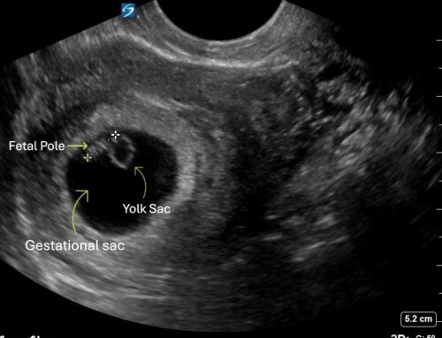
Molar pregnancy can be diagnosed with excessively high β-hcg levels and an ultrasound that shows a classic “snowstorm” or “bunches of grapes” finding.
-
Consider gestational trophoblastic disease in any patient with signs and symptoms of preeclampsia prior to 20 weeks gestation.
- Cavaliere A, Ermito S, Dinatale A, Pedata R. Management of molar pregnancy. J Prenat Med. 2009 Jan;3(1):15-7. PMID: 22439034; PMCID: PMC3279094.
- Soper, John T. MD. Gestational Trophoblastic Disease: Current Evaluation and Management. Obstetrics & Gynecology 137(2):p 355-370, February 2021. | DOI: 10.1097/AOG.0000000000004240

Krishani Patel, MD
Thomas Jefferson University Hospital
Latest posts by Krishani Patel, MD (see all)
- SAEM Clinical Images Series: Bilateral Leg Swelling with a Uterine Twist - February 6, 2026

