

The patient is a 30-year-old female with no past medical history who presents to the Emergency Department with 2 months of non-healing ulcers on multiple parts of her body. She reports getting bitten by flies while traveling in wooded trails from Venezuela through Mexico. She reports the bites started as small scabs that have since enlarged, but they are non-painful or pruritic. She has ulcerative lesions on her left hand, right arm, back, and gluteal areas. She has taken multiple antibiotics from a doctor in Mexico including clindamycin, ceftriaxone, nitrofurantoin, flagyl, and doxycycline. She denies any fevers, chills, nausea, vomiting, weight loss, or night sweats, but given the persistence of the lesions, she comes in for evaluation.


Vitals: BP 143/91 HR 60 R 17 T 98.4 O2sat 100% room air.
General: Well-appearing, breast-feeding her child.
HEENT: Oropharynx is clear, moist mucous membranes, nares clear.
Cardiovascular: Regular rate and rhythm, no murmur.
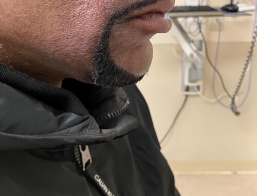
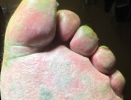
Skin: There are multiple lesions as shown in the images provided. These
are annular ulcerated pink plaques with erythematous indurated borders
and are located diffusely. The larger lesion shown is on her right arm and
is newer than the other lesions shown on her hand and trunk.
CBC: WBC: 7.6 Hgb 12.2
CRP: 0.3
Hep C/HIV/syphilis/GCCT: negative
This patient has cutaneous leishmaniasis.
Cutaneous leishmaniasis (CL) is caused by the protozoan parasite Leishmania and is transmitted through the bite of an infected female sandfly. CL is commonly diagnosed in travelers and immigrants who are susceptible to exposure. The lesions of CL usually begin as small erythematous papules that increases in size and eventually ulcerate and crust over. Lesions generally have distinct borders that are raised and erythematous. The diagnosis can be made based on travel history, lesion appearance, skin biopsy, and serology. Without treatment, the ulcers heal slowly but can leave disfiguring scars. Treatment modalities include pentamidine, amphotericin B, antimoniate, paromomycin, imiquimod, thermotherapy and cryotherapy. Visceral leishmaniasis (VL) is the most serious form of infection and is almost always fatal if untreated. With VL, the parasites directly infect organs including the spleen, liver, bone marrow, and other viscera. Common signs and symptoms of VL include fever, weight loss, fatigue, weakness, night sweats, hepatosplenomegaly, and pancytopenia.
Take-Home Points
- Cutaneous Leishmaniasis should be suspected in travelers who have been to endemic areas and present with non-healing skin ulcerations. Visceral Leishmaniasis is the most severe form of disease and carries an extremely high mortality rate if untreated.
- The vector for this disease is the sandfly; diagnosis is made through skin biopsy and direct microscopy of the parasite.
1. Eiras DP, Kirkman LA, Murray HW. Cutaneous Leishmaniasis: Current Treatment Practices in the USA for Returning Travelers. Curr Treat Options Infect Dis. 2015;7(1):52-62. doi:10.1007/s40506-015-0038-4
2. Chappuis, F., Sundar, S., Hailu, A. et al. Visceral leishmaniasis: what are the needs for diagnosis, treatment and control?. Nat Rev Microbiol 5, 873–882 (2007). https://doi.org/10.1038/nrmicro1748

Jose Gomez, MD
Emergency Medicine
UT Southwestern Medical Center
Latest posts by Jose Gomez, MD (see all)
- SAEM Clinical Images Series: Tropical Rash - October 3, 2025

Mihir Patel, MD
Emergency Medicine
UT Southwestern Medical Center
Latest posts by Mihir Patel, MD (see all)
- SAEM Clinical Images Series: Tropical Rash - October 3, 2025

Rebecca Kernen, MD
Emergency Medicine
UT Southwestern Medical Center
Latest posts by Rebecca Kernen, MD (see all)
- SAEM Clinical Images Series: Tropical Rash - October 3, 2025

Joshua Kern, MD
Assistant Professor
UT Southwestern Medical Center
Latest posts by Joshua Kern, MD (see all)
- SAEM Clinical Images Series: Tropical Rash - October 3, 2025
- SAEM Clinical Images Series: What’s Coming Out of Your Eye? - September 13, 2024
