
 The volume of women presenting to the emergency department (ED) with newly diagnosed first-trimester pregnancies and suspected ectopic pregnancies sometimes seems like an infinitely growing number. As ED physicians, proper identification of an intrauterine pregnancy (IUP) in these patients is of paramount importance and the initial imaging test of choice for many has become bedside point-of-care ultrasound (POCUS).
Value of ultrasound (POCUS) in first trimester pregnancy
The selection of POCUS as the initial imaging test compared to a radiology pelvic ultrasound (US) led to a 120-minute reduction in average ED length of stay in one single center randomized study.1 Typically these POCUS exams are performed using a standard low-frequency, transabdominal curvilinear probe. Resolution of the low-frequency curvilinear transducer limits identification of IUP if <6-7 weeks gestation, with a majority of literature estimating approximately 20% failure rate.2
![]()
Trick of the Trade: Use an ultrasound linear transducer
A recent prospective study by Tabbut et al. has shown a clinically significant decrease in the need for radiology performed transvaginal US when the high-frequency linear probe is used for detection of early IUP not seen with the curvilinear transducer.3 Briefly, here is what they found in the 81 patients studied:
- 27/81 patients did NOT have an IUP visualized with the curvilinear probe using POCUS.
- 9/27 had an IUP visualized with the high-frequency linear probe on POCUS.
- 18/27 did not visualize an IUP.
- 3/18 had an IUP seen on a radiology department performed transvaginal US study. None of the patients had an ectopic pregnancy.
Although this was a small sample size study, the authors demonstrated approximately a 10% decrease in failure rate when using high-frequency linear transducer (Figure 1).
Figure 1: Comparison of Image quality obtained with low-frequency curvilinear and linear transducer
 |
 |
Limitations of the linear transducer: The linear transducer is limited by beam penetration, with an average depth to proper identification of IUP being approximately 4 cm in the Tabbut study. Therefore image acquisition will likely be inadequate for deeper structures, a retroverted uterus, or patients using this transducer.
Technique: Identifying a first-trimester intrauterine pregnancy
Transabdominal curvilinear transducer
Scanning in the usual fashion using the curvilinear transducer, images of the uterus should be obtained in the sagittal and transverse planes (Figure 2 & 3).
Figure 2: Sagittal view of the uterus using a curvilinear transducer
Figure 3: Transverse view of the uterus using a curvilinear transducer
High frequency linear transducer
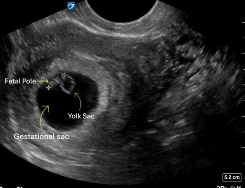
If no IUP is identified (defined as the presence of yolk sac and/or fetal pole within a gestational sac in the uterus), a transition to a linear transducer can be made. The increased frequency of the linear transducer provides improved spatial resolution up to a depth of approximately 5-6 cm depending on your transducer (Figure 4).
Figure 4: Improved resolution view of the uterus using a linear transducer

Take Home Points
The next time you may be imagining a yolk sac within a gestational sac with the curvilinear probe on POCUS, remember the magnifying glass in your back pocket – a high-frequency linear transducer.
References

Mark Favot, MD
Wayne State University School of Medicine
Co-Director, Emergency Ultrasound Fellowship
Detroit Medical Center/Wayne State University
@ten8suited
