Patwari Academy videos: Demystifiying how ECGs work

As a nice segue from the Low Risk Chest Pain videos, below is a 3-part series on Demystifying the Electrocardiogram by Dr. Rahul Patwari. It takes talent to make the complex simple.
(more…)

As a nice segue from the Low Risk Chest Pain videos, below is a 3-part series on Demystifying the Electrocardiogram by Dr. Rahul Patwari. It takes talent to make the complex simple.
(more…)
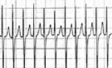
Undifferentiated tachycardias, especially when the rate is extremely fast, make it difficult to see anything other than the QRS complexes! Is there a P or flutter wave?
(more…)

The electrocardiogram can pick up all sorts of electrolyte abnormalities. The most common abnormalities revolve around high and low levels of potassium and calcium. Magnesium derangements typically have nonspecific findings. How do you keep things straight? To make things more complicated, multiple electrolyte derangements can occur at the same time, making ECG interpretation challenging.

Answer: aVR Lead
This lead can provide some unique insight into 5 different conditions:
Adapted from [1-4]
Go to ALiEM (PV) Cards for more resources.
See also:
 A patient with Parkinson’s disease presents with chest pain to your ED. Her tremors prevent you from getting a good quality EKG because of the movement artifact.
How can you eliminate this artifact? (No cheating with rocuronium.)

You always hear about it when working up syncope and sudden cardiac arrest in young patients, but it’s so easy to forget what it looks like on ECG. We so rarely see it… or DO we?!
This Paucis Verbis card on Brugada Syndrome is to help emblazon these ECG tracings in our mind, so that we don’t miss the subtle findings which place a patient at risk for sudden cardiac death. Pay special attention to Type 1, which is most specific for Brugada Syndrome.
* Update 8/2/18: Only Type 1 and Type 2 are recognized for Brugada syndrome. The type 3 pattern is likely a normal variant.
<
Adapted from [1]
Go to ALiEM (PV) Cards for more resources.
