

The patient is a 20-year-old male who presents to the Emergency Department complaining of painful and rapidly worsening swelling in the anterior neck over the last three days. The patient reports that he had a similar episode in the past for which he was prescribed antibiotics and underwent a needle aspiration procedure at the base of his mouth, which led to resolution of his symptoms. The patient reports subjective chills, change in voice, sore throat, and painful swallowing. He is able to tolerate oral secretions and denies difficulty breathing. He has no other complaints at this time. A bedside ultrasound exam and CT of the patient’s neck were subsequently performed.


Vitals: BP 147/69, HR 68, RR 18, Temp 100.6, SpO2 98% room air.
General: Nontoxic but in obvious discomfort. Able to tolerate brief
periods in a supine position. Speaking with mild stridor.
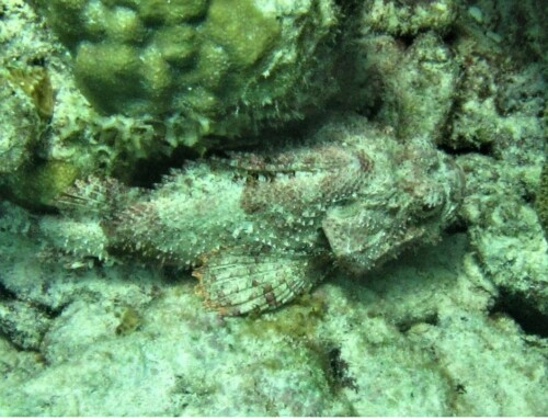
HEENT: Neck as shown. Swollen area is warm with no induration or
crepitus. Neck supple. No mastoid tenderness, normal appearing tympanic
membranes. No trismus. Normal dentition.
Respiratory: Mild biphasic stridor but normal work of breathing.
Cardiovascular: Regular rate and rhythm, no murmur.
WBC 18.0
Rapid Group A Strep swab: negative.
Monospot screen: negative
HIV antibody/antigen: negative.
Deep space infection of the neck, Ludwig’s Angina.
The patient has a plunging ranula.
Deep space infections of the neck can be categorized by the fascial layer involved: superficial, middle, and/or deep. Infections in the deep- superficial and deep-middle fascia include Ludwig’s angina, mandibular abscesses, and parotitis. These are most often caused by dental infections, and can cause compression resulting in airway compromise, thrombophlebitis, and narrowing of the great vessels of the neck. Infection of the deep layer of the cervical fascia, such as in retropharyngeal or parapharyngeal abscess, communicates directly with the mediastinum, and can rapidly progress to severe mediastinitis. This patient’s imaging shows cystic structures with no flow seen within or around the structures, and shows the fluid collection extending from the sublingual space, communicating with the submandibular space and base of the mouth. This was determined to be an infected plunging ranula.
Take-Home Points
- A simple ranula (occurs in 1 out of 5000 individuals) presents as a painless bluish saliva containing cyst visualized below the tongue.
- A “plunging” (or “diving”) ranula is a rare condition caused by direct leakage of salivary fluid from the sublingual gland at the base of the mouth into the soft tissues of the neck.
- Treatment involves needle aspiration and surgical excision. About 50% of cases recur without excision of the submandibular gland.
- Almuqamam M, Gonzalez FJ, Sharma S, et al. Deep Neck Infections. [Updated 2024 Aug 11]. In: StatPearls [Internet]. Treasure Island (FL): StatPearls Publishing; 2024 Jan-.
- Kalra V, Mirza K, Malhotra A. Plunging ranula. J Radiol Case Rep. 2011;5(6):18-24. doi: 10.3941/jrcr.v5i6.682. Epub 2011 Jun 1. PMID: 22470797; PMCID: PMC3303342.
- Olojede ACO, Ogundana OM, Emeka CI, et al. Plunging ranula: surgical management of case series and the literature review. Clin Case Rep. 2017;6(1):109-114. Published 2017 Nov 29. doi:10.1002/ccr3.1272

Peter Alsharif
Emergency Medicine
Denver Health
Latest posts by Peter Alsharif (see all)
- SAEM Clinical Images Series: An Expanding Painful Neck Mass - January 7, 2026
- SAEM Clinical Images Series: Dangerous Eye Drainage - October 23, 2023

Nhu Nguyen Le, MD
Emergency Medicine
Denver Health
Latest posts by Nhu Nguyen Le, MD (see all)
- SAEM Clinical Images Series: An Expanding Painful Neck Mass - January 7, 2026

Frederick Milgrim, MD
Emergency Medicine
Denver Health
Latest posts by Frederick Milgrim, MD (see all)
- SAEM Clinical Images Series: An Expanding Painful Neck Mass - January 7, 2026

Linda Qui, MD
Department of Emergency Medicine
Rutgers New Jersey Medical School
Latest posts by Linda Qui, MD (see all)
- SAEM Clinical Images Series: An Expanding Painful Neck Mass - January 7, 2026