

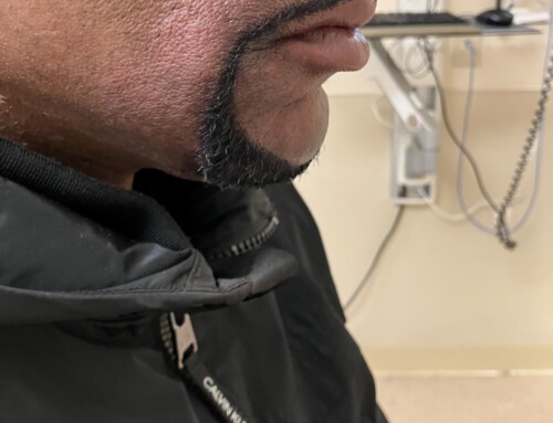
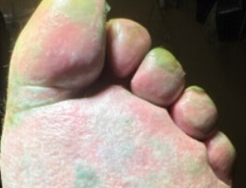
A 76-year-old female presented with a lingering cough and an oral lesion to the left lower cheek. She reported ten days of improving flu-like symptoms but had a persistent cough and nasal congestion. On the day of presentation, she developed a painful, intermittently bleeding “blood blister” to the left lower cheek that had increased in size, as well as new red spots on her arms and legs. She reported no recent trauma or history of similar lesions in the past.

Jeffrey Cheah, MD
Resident Physician
Boston Medical Center
Boston Medical Center
Latest posts by Jeffrey Cheah, MD (see all)
- SAEM Clinical Images Series: A Blistery Mystery - October 11, 2024

Bethanne Bartscherer, MD
Resident Physician
Boston Medical Center
Boston Medical Center
Latest posts by Bethanne Bartscherer, MD (see all)
- SAEM Clinical Images Series: A Blistery Mystery - October 11, 2024

Anna Fang, MD
Resident Physician
Department of Emergency Medicine
Boston Medical Center
Department of Emergency Medicine
Boston Medical Center
Latest posts by Anna Fang, MD (see all)
- SAEM Clinical Images Series: A Blistery Mystery - October 11, 2024
- SAEM Clinical Images Series: A Painful Swollen Digit - August 21, 2023

Andrew Mittelman, MD
Assistant Professor of Emergency Medicine
Boston Medical Center
Latest posts by Andrew Mittelman, MD (see all)
- SAEM Clinical Images Series: A Grain of Sand… or Something More Sinister? - February 27, 2026
- SAEM Clinical Images Series: A Pedunculated Bone to Pick - January 12, 2026
- SAEM Clinical Images Series: Tangled in the Toilet - October 31, 2025
