

A female in her 50s with a past medical history of coronary artery disease, pacemaker placement, hypertension, and ESRD presented to the emergency department with the chief complaint of missed dialysis, breast engorgement, and an increase in vascularity in her chest and abdomen. The patient reported an increase in breast swelling and increased vascularity in her belly over the past three months. Additionally, she woke up short of breath on the morning of presentation and reported dyspnea at rest. She denied chest pain, diaphoresis, breast pain, fever, rash, trauma to the breasts, or drainage.


Vitals: T 36.9°C; HR 105; BP 109/74; RR 20; O2 sat 97% on nasal canula @ 3L
Neck: JVD
Lungs: Bilateral crackles
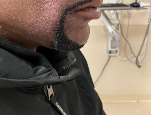
Chest and abdomen: Increased vascularity
Breast: Bilateral breast swelling and redness
Lower extremity: Bilateral pitting edema and varicose veins
Basic metabolic panel (BMP): K 6.9; Cr 9.53
Brain natriuretic peptide (BNP): >35,000
Troponin I: 0.1
DDX: Inflammatory carcinoma, mastitis, superior vena cava syndrome, portal hypertension, pulmonary hypertension, pulmonary embolism.
Superior vena cava (SVC) syndrome results from any condition that leads to obstruction of blood flow through the SVC. Our case was caused by complete occlusion from a thrombus and the patient presented with bilateral breast swelling, skin changes (peau d’orange), and an increase in vascularity in the abdomen and chest (caput medusa). Breast tissue largely drains into the axillary veins, and more proximally into the subclavian veins. Due to occlusion of the SVC, a complete backup of venous flow occurs, resulting in all of the noted collateral hypervascularity. Often SVC occlusion is caused by malignancy obstructing the superior vena cava or invading the vein.
The CTA demonstrates occlusion of the superior vena cava. There are multiple varices in the chest wall and the imaged upper abdominal wall. There is also diffuse subcutaneous edema with diffuse soft tissue swelling and skin thickening of the bilateral breasts.
Take-Home Points
- Consider superior vena cava occlusion in patients undergoing hemodialysis who present with the above physical exam findings.
- Consider occult malignancy as the source or cause of thrombosis.
- Be sure to fully expose your patient when appropriate and keep your differential broad.
- Corduff N, Rozen WM, Taylor GI. The superficial venous drainage of the breast: a clinical study and implications for breast reduction surgery. J Plast Reconstr Aesthet Surg. 2010 May;63(5):809-13. doi: 10.1016/j.bjps.2009.02.055. Epub 2009 Apr 3. PMID: 19345164.
- Friedman T, Quencer KB, Kishore SA, Winokur RS, Madoff DC. Malignant Venous Obstruction: Superior Vena Cava Syndrome and Beyond. Semin Intervent Radiol. 2017 Dec;34(4):398-408. doi: 10.1055/s-0037-1608863. Epub 2017 Dec 14. PMID: 29249864; PMCID: PMC5730434.

Martin Morales Cruz, MD
Department of Emergency Medicine
UCF HCA GME Consortium EM Residency
Osceola Regional Medical Center
Latest posts by Martin Morales Cruz, MD (see all)
- SAEM Clinical Images Series: Breast Swelling - August 29, 2022

Mark Rivera-Morales, MD
Department of Emergency Medicine
UCF HCA GME Consortium EM Residency
Osceola Regional Medical Center
Latest posts by Mark Rivera-Morales, MD (see all)
- SAEM Clinical Images Series: Breast Swelling - August 29, 2022
- Ultrasound for the Win! 18M with Acute Shoulder Injury #US4TW - November 11, 2020

Larissa Dub, MD
Department of Emergency Medicine
Osceola Regional Medical Center
Latest posts by Larissa Dub, MD (see all)
- SAEM Clinical Images Series: Breast Swelling - August 29, 2022
- SAEM Clinical Image Series: Juvenile Snake Bite - September 7, 2020
