

The patient is an 81-year-old female with a history of asthma and hypertension who presents to the Emergency Department with right-sided abdominal swelling for five days. Five days ago, the right side of her abdomen appeared to protrude more than the left. This protrusion then increased over the next 2-3 days. The patient was diagnosed with shingles to the right lower abdomen earlier that month, but her rash has now nearly resolved. She continues to have “electric” pain in the region of the prior shingles infection. She denies any fevers, abdominal trauma, vomiting, or changes in bowel or bladder habits. She has never had anything like this before.


Vitals: All vital signs are normal.
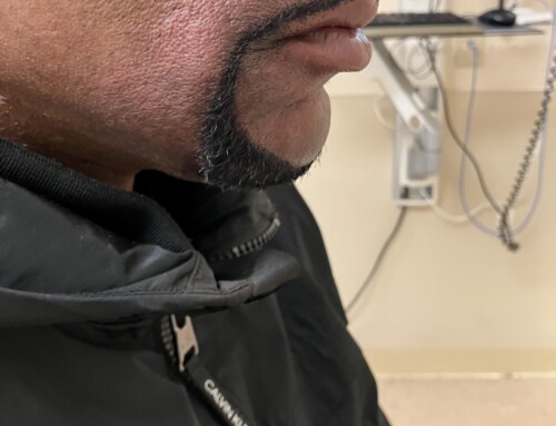
Abdomen: See image provided. There is a firm unilateral distention of the right lower abdomen without shifting dullness or fluid wave. No palpable masses are present. There is moderate tenderness over the protruding region but no rebound or guarding. Bowel sounds are present.
Skin: See image provided. Moderate tenderness to palpation over region.
CBC and CMP are unremarkable.
CT scan of the abdomen and pelvis: No evidence of acute abnormality. Normal appendix. Moderate stool burden.
This patient has a Zoster pseudohernia.
Varicella-Zoster Virus (Shingles).
Zoster pseudohernia (ZP) occurs when the zoster infection infiltrates a posterior thoracic dermatome, affecting the spinal nerve roots responsible for the motor function of the abdominal wall. ZP typically presents with a rapidly progressive unilateral outpouching of the abdomen, giving a hernia-like appearance, but with intact abdominal wall musculature. In most cases, the classic Herpes Zoster rash precedes ZP, however in up to 10% of patients ZP may be the first presenting sign of zoster. Although uncommon, the symptoms are often distressing to patients, with many presenting to emergency departments or surgical offices for initial evaluation. The diagnosis is clinical and is based on a history of zoster infection or classic zoster symptoms and lack of findings suggesting alternate pathology. Abdominal CT or other imaging is recommended to exclude tumors, true hernias, free fluid, or other possible causes of abdominal distention. Electromyography (EMG) can be used to support diagnosis and will typically be abnormal due to the dysfunction of the abdominal wall musculature. Complete recovery occurs in 70-80% of patients within about 4-5 months.
Take-Home Points
Zoster Pseudohernia is a rare presentation of herpes zoster infection resulting in dysfunction of the abdominal wall musculature.
There is no specific treatment with most cases fully resolving within several months to one year.
- Chernev I, Dado D. Segmental zoster abdominal paresis (zoster pseudohernia): a review of the literature. PM R. 2013 Sep;5(9):786-90. doi: 10.1016/j.pmrj.2013.05.013. PMID: 24054853.
- Yoo J, Koo T, Park E, Jo M, Kim MS, Jue MS. Abdominal pseudohernia caused by herpes zoster: 3 case reports and a review of the literature. JAAD Case Rep. 2019 Aug 5;5(8):729-732. doi: 10.1016/j.jdcr.2019.06.019. PMID: 31440570; PMCID: PMC6698640.

Ana Hardisty, MD
Southwest Healthcare MEC
Latest posts by Ana Hardisty, MD (see all)
- SAEM Clinical Images Series: Pain, Paralysis, and Rash - January 26, 2026

Robert Steele, MD
Southwest MEC
Latest posts by Robert Steele, MD (see all)
- SAEM Clinical Images Series: Pain, Paralysis, and Rash - January 26, 2026
- SAEM Clinical Images Series: Leg Rash - November 10, 2025

