

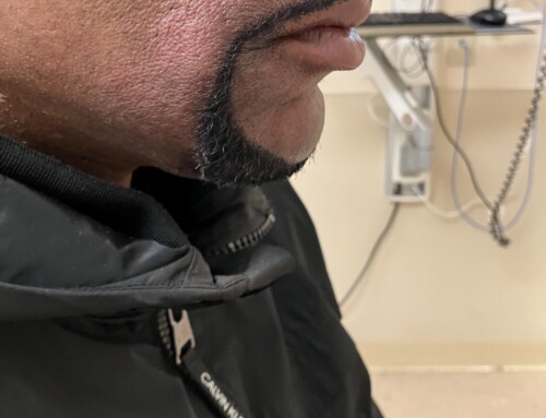
A 16-month-old male with no significant past medical history presented with a chief complaint of bleeding from the mouth. The patient’s mother looked inside his mouth and described a “black ball of flesh” near the right side of his lower gum. The mother noted that he had been more fussy than usual and appeared to have a decreased appetite over the past few days. The mother was unsure how long the lesion had been present. The mother denied any recent witnessed falls, trauma, or injury. The mother denied any recent fever, emesis, skin rashes, or lesions. She reported that the patient is an otherwise healthy child without any drug allergies or daily medications.

General: He is not in acute distress. He is well-developed.
HEENT: Head: Normocephalic and atraumatic. Nose: Nose normal. No congestion or rhinorrhea. Mouth: Mucous membranes are moist. Purple-colored flesh- appearing nodule erupting from right lower gum. Dentition is intact and well-appearing.
Pharynx: Oropharynx is clear.
Skin: Warm and dry. No other skin rashes, lesions, or abrasions.
None
An eruption cyst (EC) is a dome-shaped soft tissue lesion associated with the eruption of primary or permanent teeth. An eruption hematoma forms when the cyst fluid contains blood, often appearing blue or black.
Differential diagnosis:
- Retrocuspid papillae are small, firm, round, pink to red papules on the posterior surface of the gums, typically behind the lower canine teeth in most children. They are often bilateral.
- Parulis or “gum boil” is a soft, solitary, red papule on the gums above or below a necrotic tooth, typically forming over a fistulous tract between the abscess and gums.
- Dentigerous cyst (DC) is a well-defined area of radio-opacity that is characterized by permanent teeth that are incapable of eruption.
- Neonatal alveolar lymphangioma (NAL) is a rare, benign condition that presents with a bluish-black fluid-filled dome on the alveolar ridge surface. This condition is most often seen in black neonates.
- Oral hemangiomas are benign tumors that develop due to endothelial cell proliferation. The majority of these tumors will resolve over time and do not require treatment.
- Amalgam tattoo is a localized area of blue, gray, or black pigmentation that is caused by excess amalgam inadvertently embedded during a dental procedure.
Eruption cysts are typically asymptomatic and will not require active treatment. The majority of ECs burst spontaneously with the passage of the tooth. If the cyst is symptomatic, simple surgical excision by a dental profressional is recommended, as well as pain control with acetaminophen and ibuprofen. This procedure consists of incising the cyst roof to allow drainage of fluid and descent of the tooth.
Take-Home Points
-
Eruption cysts can be managed conservatively with pain control and anticipatory guidance.
-
If symptomatic, patients with eruption cysts should be referred to a dental provider for further evaluation and possible surgical excision.
-
If the eruption cyst does not resolve within two weeks, the patient should be evaluated for other causes.
-
Dhawan, Preeti, et al. “Eruption cysts: A series of two cases.” Dental Research Journal, vol. 9, no. 5, 2012, p. 647, https://doi.org/10.4103/1735-3327.104889.
-
Keels, Martha Ann. “Soft Tissue Lesions of the Oral Cavity in Children.” UpToDate, www.uptodate.com/contents/soft-tissue-lesions-of-the-oral-cavity-in-children/print. Accessed 28 Dec. 2023.
-
Sen-Tunc, E, et al. “Eruption cysts: A series of 66 cases with clinical features.” Medicina Oral Patología Oral y Cirugia Bucal, 2017, pp. 0–0, https://doi.org/10.4317/medoral.21499.

Madison Allen, MD
Medical College of Wisconsin
Latest posts by Madison Allen, MD (see all)
- SAEM Clinical Images Series: What is in my Child’s Mouth? - March 17, 2025

Wendi Wendt, MD
Pediatric Emergency Medicine
Medical College of Wisconsin
Latest posts by Wendi Wendt, MD (see all)
- SAEM Clinical Images Series: A Mucous Membrane Mystery - February 20, 2026
- SAEM Clinical Images Series: Can I Snooze on This Bruise? - January 9, 2026
- SAEM Clinical Images Series: What is in my Child’s Mouth? - March 17, 2025
