Teaching internationally: More than just a language barrier

JoshiUltrasound1I recently traveled to San Salvador to help teach a pediatric and adult ultrasound course. The course was well received and it was wonderful traveling around San Salvador.

I wanted to share some of our experiences, and discuss some challenges to educating internationally. More importantly, I want to engage you, the readers to share some of your experiences when educating internationally as well.

(more…)

By |2016-11-11T18:43:05-08:00Dec 7, 2012|Medical Education, Ultrasound|

Trick of the Trade: Ultrasound-guided injection for shoulder dislocation

ShoulderDislocation

Who loves relocating shoulder dislocations as much as I do? I know you do.

Often patients undergo procedural sedation in order to achieve adequate pain control and muscle relaxation. Alternatively or adjunctively, you can inject the shoulder joint with an anesthetic. Personally, I have had variable effectiveness with this technique. In cases of inadequate pain control, I always wonder if I was actually in the joint.

How can you improve your success rate in injecting into glenohumeral joint injection?

(more…)

By |2019-01-28T22:35:26-08:00Nov 8, 2011|Orthopedic, Tricks of the Trade, Ultrasound|

Paucis Verbis: Diagnostic testing tips for acute abdominal pain

acute abdominal pain

In the most recent EM Clinics of North America publication, Dr. Panebianco et al. discussed the evidence behind diagnostic tests for acute abdominal pain. There were some really great teaching points in this broad-reaching topic.

My favorite pearl: A 3-way acute abdominal series is too insensitive to rule-out any major acute causes of abdominal pain with confidence. So stop ordering them routinely. If you are worried about a perforated viscus, order an upright chest x-ray instead — more accurate and less radiation.


Adapted from [1]
Go to ALiEM (PV) Cards for more resources.

Reference

  1. Panebianco N, Jahnes K, Mills A. Imaging and laboratory testing in acute abdominal pain. Emerg Med Clin North Am. 2011;29(2):175-93, vii. [PubMed]
By |2021-10-12T16:23:35-07:00Jul 22, 2011|ALiEM Cards, Gastrointestinal, Radiology|

Tricks of the Trade: Underwater ultrasonography

Basketweaving

I’ve heard of underwater basketweaving, but underwater ultrasonography?

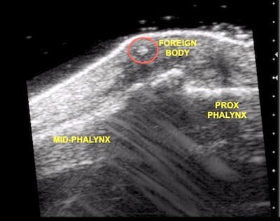
Bedside ultrasonography is a great tool to help find small foreign bodies. Commonly foreign bodies get lodged superficially in the patient’s extremities. Because superficial structures (<1 cm deep) are difficult to visualize on ultrasound, you should apply a really generous, thick layer of ultrasound gel to create some distance. Alternatively, you can add a step-off pad, such as a bag of saline or fluid-filled glove, to place between the patient’s skin and transducer. What’s a quicker and easier way to create some distance yet preserve image quality?

Trick of the Trade

Submerse both the body part and the ultrasound transducer under water.

ultrasonography water bath

For this “bath water technique”, start by holding the transducer perpendicular to the wound and about 1 cm away from the skin. You can adjust the distance to optimize the image quality.

FB_Hand

FB_Hand2

Thanks to Andy at Emergency Medicine Ireland blog for these 2 ultrasound images! 

This submersion technique has been published in American Journal of EM in 2004 as a painless alternative to gel or a step-off pad, because the transducer does not need to apply any pressure on the patient’s wound.

Reference
Blaivas M, Lyon M, Brannam L, Duggal S, Sierzenski P. Water bath evaluation technique for emergency ultrasound of painful superficial structures. Amer J Emerg Med. 2004; 22(7), 589-93 PMID: 15666267

By |2020-04-21T12:30:07-07:00Jul 20, 2011|Tricks of the Trade, Ultrasound|
Go to Top