Paucis Verbis: Head CT clinical decision rules in trauma
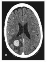
HeadCTbleedThe ideal clinical decision tool has a sensitivity and specificity of 100%.

You need a high sensitivity to be sure that your negative result indeed predicts a true negative. That means if your clinical decision tool suggests that you don’t need to get a head CT, then your head CT would have been normal.

On the flip side, this realistically means there is a low-moderate specificity. That means a clinical decision tool with at least 1 positive criterion does not always mean that there will be an abnormal finding on head CT.

There are 3 major clinical decision rules that I’ve heard tossed around in the literature:

  • Canadian CT Head Rules (CCHR)
  • New Orleans Criteria (NOC)
  • National Emergency X-Radiography Utilization Study (NEXUS)-II

There is no perfect tool.

Take a look at these decision rules and their inclusion criteria.

  • The CCHR included patients with GCS 13-15. The NOC initially enrolled only patients with a GCS of 15.
  • All factor in age (≥65 years for CCHR and NEXUS-II; ≥60 years for NOC).
  • Interestingly only the CCHR, for better or worse, take into account mechanism of injury. I’m not sure I would obtain a head CT on a pedestrian with a graze wound on the foot from a slow-moving vehicle.

Which do you use? I use a combination of all 3 and my clinical gestalt.

PV Card: Head CT in Trauma – Clinical Decision Tools


Go to ALiEM (PV) Cards for more resources.

By |2021-10-15T10:59:57-07:00May 13, 2011|ALiEM Cards, Radiology, Trauma|

Tricks of the Trade: Ultrasound workshop setup

 
Ultrasound

Have you ever been to an ultrasound workshop where each small group of attendees huddles around the small ultrasound display? Personally I think the 3 people closest to the display really see the images well. This tends to exclude the other participants.

Last week, I hosted (my first!) ultrasound workshop for the UCSF Alumni CME Conference where I showed peri-retired UCSF alumni from various specialties about the future of bedside ultrasonography. I equated it to the 21st century stethoscope. Thanks to my star team of ultrasonographers: Dr. Asaravala, Flores, Miss, Lenaghan, and Wilson.

(more…)

By |2016-11-11T18:54:22-08:00May 11, 2011|Tricks of the Trade, Ultrasound|

Trick of the Trade: Oblique CXR to look for pneumothorax


PneumothoraxCTSupine chest xrays have an extremely low sensitivity (12-24%) in detecting pneumothoraces. Because a pneumothorax layers anteriorly on an AP CXR film, the xray beam is perpendicular rather than tangential to the pneumothorax edge. This makes visualizing a small to moderate sized pneumothorax extremely difficult. So you are left to look for indirect signs such as a deep sulcus at the costophrenic angle or subcutaneous air. I’m often surprised at how large of a pneumothorax can be missed on CXR based on CT imaging. The image to the right shows a large left-sided pneumothorax on CT.

What if you have an equivocal bedside ultrasound result in looking for a pneumothorax, and the patient is too unstable to go to CT?

 
By |2019-02-19T18:08:06-08:00Apr 13, 2011|Radiology, Tricks of the Trade|

Trick of the Trade: Check pupillary constriction with ultrasound

SwollenEyeIn some trauma patients with head and face trauma, you will need to check their pupillary response to light. Severe periorbital and eyelid swelling, however, make this difficult. You want to minimize multiple attempts to retract the eyelids because of the risk of a ruptured globe. What’s a minimally painful and traumatic way to check for pupillary constriction?

By |2019-01-28T22:50:28-08:00Apr 6, 2011|Ophthalmology, Tricks of the Trade, Ultrasound|

Trick of the Trade: Ultrasound-guided supraclavicular central line

SupraclavicularPositionsmEmergency physicians are procedural experts in central venous access. The subclavian vein is the best site for such access, because it has been shown to have the lowest rate of iatrogenic infections and deep venous clots

Bedside ultrasonography has really revolutionized how we obtain vascular access over the past 10 years. Identifying the subclavian vein using ultrasonography, however, is still technically challenging. The vein is located just posterior to the clavicle, which often gets in the way of the linear transducer. 

By |2016-11-11T19:00:20-08:00Nov 10, 2010|Tricks of the Trade, Ultrasound|

Trick of the trade: I got ultrasound gel in my eye!

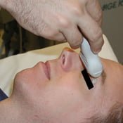
OcularUltrasoundProbeBedside ultrasonography is increasingly being used in the ED to examine the eye. For instance, it can be used to detect a retinal detachment, vitreous hemorrhage, and high intracranial pressure. The technique involves applying ultrasound gel on the patient’s closed eyelid. A generous amount of gel should be used to minimize the amount of direct pressure applied on the patient’s eye by the ultrasound probe.

Sometimes, however, no matter how careful you and the patient are, some gel accidentally contacts the eye itself.

(more…)

By |2019-01-28T23:23:16-08:00Sep 15, 2010|Tricks of the Trade, Ultrasound|
Go to Top