

What anticoagulant medication can cause these skin changes?
- Apixaban
- Heparin
- Rivaroxaban
- Warfarin
[Image courtesy of Herbert Fred, MD and Hendrik van Dijk via Wikimedia Commons]

The American College of Medical Toxicology hosts this Toxicology Visual Pearls series. The post was peer reviewed on behalf of ACMT by Drs. Howard Greller, Louise Kao, and Mark K Su.
References
- Pourdeyhimi, N., Bullard, Z. (2014). Warfarin-induced skin necrosis. Hosp Pharm. 2014;49(11):1044–1048. PMID: 25673894
- Nsaful J, Adjei YO, Dedey F, Agboadoh N, Anyigba E, Pieterson W. Warfarin-induced skin necrosis: a rare condition. Ghana Med J. 2020;54(4):269-273.PMID: 33883776
- Verhagen H. Local haemorrhage and necrosis of the skin and underlying tissues, during anti-coagulant therapy with dicumarol or dicumacyl. Acta Med Scand. 1954;148(6):453-467.PMID: 13171021
- DeFranzo AJ, Marasco P, Argenta LC. Warfarin-induced necrosis of the skin. Ann Plast Surg. 1995;34(2):203-208. PMID: 7741443
- Kakagia DD, Papanas N, Karadimas E, Polychronidis A. Warfarin-induced skin necrosis. Ann Dermatol. 2014;26(1):96-98. PMID: 24648693
- Ejzenberg D, Neusquen LP, Rolnik DL, Lozinsky AC, Piato JR. Breast necrosis induced by the use of coumadin: case report and review of literature. Einstein (Sao Paulo). 2015;13(3):417-419. PMID: 26018146
- Eby CS. Warfarin-induced skin necrosis. Hematol Oncol Clin of North Am. 1993;7(6):1291–1300. PMID: 8294318
- Chan YC, Valenti D, Mansfield AO, Stansby G. Warfarin induced skin necrosis. Br J Surg. 2000;87(3):266-272.PMID: 10718793
- Srinivasan AF, Rice L, Bartholomew JR, et al. Warfarin-induced skin necrosis and venous limb gangrene in the setting of heparin-induced thrombocytopenia. Arch Intern Med. 2004;164(1):66-70. PMID: 14718324

Natalie M. Rall, MD
Emergency Medicine Chief Resident
Carolinas Medical Center
Carolinas Medical Center
Latest posts by Natalie M. Rall, MD (see all)
- ACMT Visual Pearl: Necrosis in the Name of Anticoagulation - November 4, 2025

Erik Fisher, MD
Associate Program Director, Medical Toxicology Fellowship
Atrium Health Carolinas Medical Center and Levine Children's Hospital
Clinical Assistant Professor of Emergency Medicine
Wake Forest School of Medicine
Atrium Health Carolinas Medical Center and Levine Children's Hospital
Clinical Assistant Professor of Emergency Medicine
Wake Forest School of Medicine
Latest posts by Erik Fisher, MD (see all)
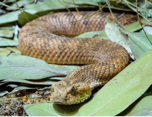
- ACMT Toxicology Visual Pearl – The Fang and the Furious - June 30, 2026
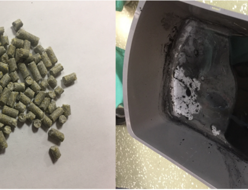
- ACMT Toxicology Visual Pearl: Delayed Skin Burn - May 5, 2026
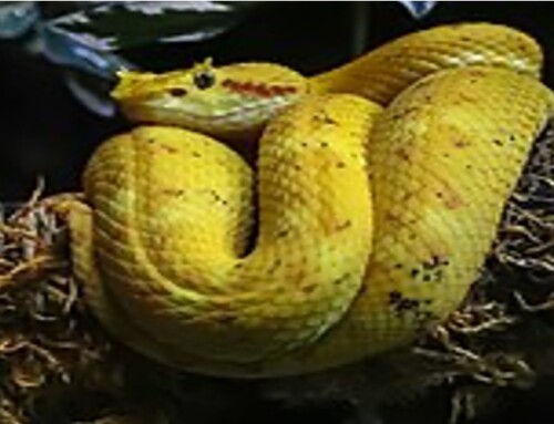
- ACMT Toxicology Visual Pearl: Sleeping with the Fishes - February 24, 2026