Paucis Verbis: Spinal epidural abscess

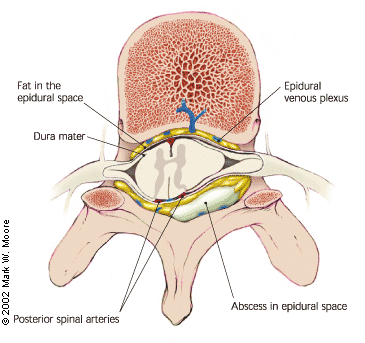
Spinal epidural abscess anatomy illustrationOne of the most challenging diagnoses to make is that of a spinal epidural abscess (SEA), especially if you work in an Emergency Department which cares for many IV drug users and HIV patients. There’s never before been a published diagnostic guideline or algorithm which helps you with risk-stratification.

In the Journal of Neurosurgical Spine, a diagnostic guideline was prospectively evaluated on a small population (n=31) as compared to historical controls (n=55). They found that an ESR test had a sensitivity of 100% if a patient had at least 1 risk factor for SEA. A CRP test was much less helpful.

Not a practical algorithm

Unfortunately, they didn’t study the utilization rate of the MRI scanner with this guideline. Are they getting better results (fewer diagnostic delays and fewer cases of patients later in their clinical course) because they are just MRI-scanning more people? Almost everyone in my ED with back pain would fall into the Urgent/Emergent MRI box…  I’m not a fan of this algorithm.

Regardless, this algorithm may help you in shaping your diagnostic decision and medical decision making documentation.

PV Card: Spinal Epidural Abscess


Adapted from [1]
Go to ALiEM (PV) Cards for more resources.

Reference

  1. Davis D, Salazar A, Chan T, Vilke G. Prospective evaluation of a clinical decision guideline to diagnose spinal epidural abscess in patients who present to the emergency department with spine pain. J Neurosurg Spine. 2011;14(6):765-770. [PubMed]
By |2021-10-12T16:17:13-07:00Aug 5, 2011|ALiEM Cards, Infectious Disease, Neurology|

Paucis Verbis: Fibrinolytics for Acute Pulmonary Embolism

Pulmonary embolism fibrinolytics

When would you give fibrinolytics for a Pulmonary Embolism?

This Paucis Verbis card summarizes recommendations found in Circulation’s recently published Scientific Statement from the American Heart Association. Although it is rare to give fibrinolytics for a pulmonary embolism (PE) in the Emergency Department, it is important to remember when lytics are indicated.

PV Card: Fibrinolysis for Acute Pulmonary Embolism


Adapted from [1]
Go to ALiEM (PV) Cards for more resources.

Reference

  1. Jaff M, McMurtry M, Archer S, et al. Management of massive and submassive pulmonary embolism, iliofemoral deep vein thrombosis, and chronic thromboembolic pulmonary hypertension: a scientific statement from the American Heart Association. Circulation. 2011;123(16):1788-1830. [PubMed]
By |2021-10-12T16:21:19-07:00Jul 29, 2011|ALiEM Cards, Cardiovascular, Pulmonary|

Trick of the Trade: Making a beanie hat

Scalp lacerations are a common condition in the Emergency Department. Some require no bandage over once the injury is repaired. Because the scalp is so vascular, others require a pressure dressing over the site to minimize hematoma formation.

How do you bandage these patients? It is difficult to secure any wrap or square gauze over the site, because the head is round and the hair is slippery.

(more…)

By |2019-01-28T22:36:18-08:00Jul 27, 2011|Tricks of the Trade|

Paucis Verbis: Diagnostic testing tips for acute abdominal pain

acute abdominal pain

In the most recent EM Clinics of North America publication, Dr. Panebianco et al. discussed the evidence behind diagnostic tests for acute abdominal pain. There were some really great teaching points in this broad-reaching topic.

My favorite pearl: A 3-way acute abdominal series is too insensitive to rule-out any major acute causes of abdominal pain with confidence. So stop ordering them routinely. If you are worried about a perforated viscus, order an upright chest x-ray instead — more accurate and less radiation.


Adapted from [1]
Go to ALiEM (PV) Cards for more resources.

Reference

  1. Panebianco N, Jahnes K, Mills A. Imaging and laboratory testing in acute abdominal pain. Emerg Med Clin North Am. 2011;29(2):175-93, vii. [PubMed]
By |2021-10-12T16:23:35-07:00Jul 22, 2011|ALiEM Cards, Gastrointestinal, Radiology|

Tricks of the Trade: Underwater ultrasonography

Basketweaving

I’ve heard of underwater basketweaving, but underwater ultrasonography?

Bedside ultrasonography is a great tool to help find small foreign bodies. Commonly foreign bodies get lodged superficially in the patient’s extremities. Because superficial structures (<1 cm deep) are difficult to visualize on ultrasound, you should apply a really generous, thick layer of ultrasound gel to create some distance. Alternatively, you can add a step-off pad, such as a bag of saline or fluid-filled glove, to place between the patient’s skin and transducer. What’s a quicker and easier way to create some distance yet preserve image quality?

Trick of the Trade

Submerse both the body part and the ultrasound transducer under water.

ultrasonography water bath

For this “bath water technique”, start by holding the transducer perpendicular to the wound and about 1 cm away from the skin. You can adjust the distance to optimize the image quality.

FB_Hand

FB_Hand2

Thanks to Andy at Emergency Medicine Ireland blog for these 2 ultrasound images! 

This submersion technique has been published in American Journal of EM in 2004 as a painless alternative to gel or a step-off pad, because the transducer does not need to apply any pressure on the patient’s wound.

Reference
Blaivas M, Lyon M, Brannam L, Duggal S, Sierzenski P. Water bath evaluation technique for emergency ultrasound of painful superficial structures. Amer J Emerg Med. 2004; 22(7), 589-93 PMID: 15666267

By |2026-06-16T16:03:08-07:00Jul 20, 2011|Tricks of the Trade, Ultrasound|

Paucis Verbis: NSAIDS and upper GI bleeds

NSAIDs gi bleed

Do no harm.

We so often recommend and give NSAIDs to patients for various painful conditions. We also commonly administer ketorolac (toradol) in the ED, because it works so amazingly well for renal colic. When giving various NSAIDs, what is the relative risk (RR) for an upper GI bleed or perforation in the first year?

Ketorolac has the highest upper GI complication RR (14.54) for all of the studied NSAIDs. Compare this with the overall risk of traditional COX-1 NSAIDS (RR=4.5) and COX-2 inhibitors (RR=1.88). So before giving ketorolac, first check that patients don’t have a history of a GI bleed or peptic ulcer.

PV Card: NSAIDs and Upper GI Bleed


Adapted from [1]
Go to ALiEM (PV) Cards for more resources.

Reference

  1. Massó G, Patrignani P, Tacconelli S, García R. Variability among nonsteroidal antiinflammatory drugs in risk of upper gastrointestinal bleeding. Arthritis Rheum. 2010;62(6):1592-1601. [PubMed]
By |2021-10-12T16:27:37-07:00Jul 15, 2011|ALiEM Cards, Gastrointestinal, Tox & Medications|
Go to Top