
SplintER Series: To Immobilize or Not to Immobilize: That is the Question

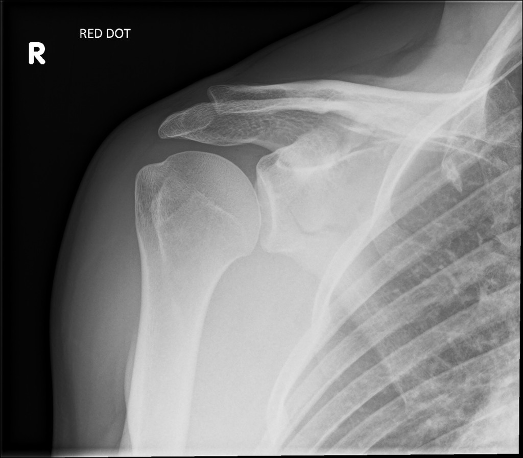
A patient presents to the Emergency Department after sustaining a twisting knee injury while skiing. She felt a pop and was unable to bear weight afterward secondary to pain and a feeling of instability. Shortly after the injury, she noted increased swelling and pain. On examination, she has a moderate effusion and a positive Lachman test. An x-ray was obtained and is shown above (Image 1. Case courtesy of Mikael Häggström, M.D. – Author info – Reusing images, CC0, via Wikimedia Commons).


 A 76-year-old female with a history of HTN, TIA, CAD, left CEA, and CKD presented to the emergency department for evaluation of neck bruising and swelling. The patient stated that the night before, she was eating popcorn and choked on a kernel. She states that she coughed to clear her throat and shortly after she developed swelling and bruising to the left side of her neck, which has progressively gotten worse. The patient has a remote history of left carotid endarterectomy and was concerned that her symptoms could be related to the prior surgery. On examination, she had ecchymosis and a hematoma/mass to the left side of her neck without palpable thrill or bruit. A well-healed CEA scar was noted. A CTA of the neck was obtained to determine the source of the ecchymosis/hematoma. What is the diagnosis?
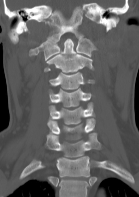


