Trick of the Trade: Hip dislocation Part II
 As a followup to the blog on the Captain Morgan technique for hip dislocations, I’d like to throw out another similar technique that also does NOT involve climbing up on the gurney.



As a followup to the blog on the Captain Morgan technique for hip dislocations, I’d like to throw out another similar technique that also does NOT involve climbing up on the gurney.
Relocation of a hip joint is often quite a sight to see in the ED. A commonly taught technique is the Allis maneuver (watch the first 45 seconds of the above video from the Medical College of Georgia). It has always seemed a bit precarious to me having someone stand on the patient’s bed.
(more…)

Who loves relocating shoulder dislocations as much as I do? I know you do.
Often patients undergo procedural sedation in order to achieve adequate pain control and muscle relaxation. Alternatively or adjunctively, you can inject the shoulder joint with an anesthetic. Personally, I have had variable effectiveness with this technique. In cases of inadequate pain control, I always wonder if I was actually in the joint.
How can you improve your success rate in injecting into glenohumeral joint injection?
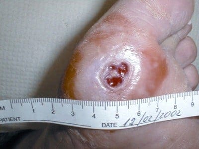
We sometimes see diabetic patients in the ED for a worsening foot ulcer. Sometimes it’s the chief complaint. Other times, however, you just notice it on physical exam. So, be sure you examine the feet of your diabetic patients. Occasionally, you’ll be surprised by what you find.
Several questions come up with diabetic foot ulcers:
Thanks to JAMA‘s Clinical Rational Examination series, there is a systematic review of diabetic leg ulcers and osteomyelitis. Here are the highlights:
Adapted from [1]
Go to ALiEM (PV) Cards for more resources.
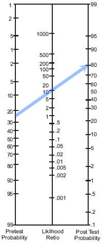
Below is the Fagan nomogram to help you plot out your post-test probability based on your likelihood ratios. The example given is if your pretest probability is 25% and your LR is 10. Your post-test probability would be 80%.
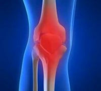
A 55 year old woman presents with rheumatoid arthritis presents with monoarticular joint pain in her left knee for the past 3 days. She has a low-grade fever of 100.2 F and a significantly warm and tender knee. “It feels different than my RA flare.”
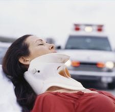
“Distracting injury” is a frequent cited reason for imaging the cervical spine in blunt trauma patients, per the NEXUS study. In the Journal of Trauma in 2005 and 2011, studies aimed to narrow the definition of “distracting injury”. Although both are studies at different sites, both conclude the same:
So let’s say you are caring for a non-intoxicated motor vehicle crash patient with an isolated tibia fracture (i.e. a “long bone fracture”), no chest injuries, and no neck pain/tenderness. Per the NEXUS criteria, you might consider this patient to have a “distracting injury” because of the long bone fracture. Instead, the literature now supports your clinically clearing the cervical spine without imaging.
Wait, let’s rethink this. Does this mean that you should get cervical spine imaging for ALL blunt trauma patients with ANY chest wall tenderness?! NO. That’s just crazy. You should still factor in the mechanism of injury, severity of pain, and your clinical gestalt.
So for me, these “distracting injury” studies are helpful such that:
Go to ALiEM (PV) Cards for more resources.
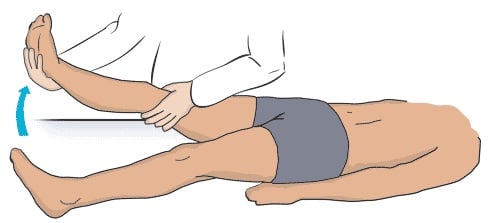 A 35 year old man presents with low back pain which radiates down his right leg to the level of the knee. Is this sciatica?
Low back pain is one of the most common chief complaints that we see in the Emergency Department. In addition to the examination of the back and distal neurovascular function, we also need to test for evidence of a radiculopathy (compression or inflammation of a nerve root typically from a herniated disk). Because most disk herniations occur at the L4-L5 and L5-S1 level, you should test for irritation of the L4-S1 nerve roots. This is the sciatic nerve.