


Adenosine in reactive airway disease
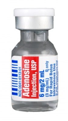
A 44-year old woman presents via EMS with a chief complaint of a racing heartbeat. She is placed on a cardiac monitor, which displays a heart rate of 192, and a subsequent EKG reveals she is in SVT. She also complains of chest discomfort and shortness of breath. Her blood pressure is stable, and you decide to treat her with adenosine. As you take a more thorough past medical history, you learn your patient has a history of asthma. One of the EM residents mentions that he thought adenosine should not be given to patients with reactive airway disease.

 Welcome to the seventh ALiEM
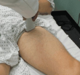 Deep vein thrombosis (DVT) is always a consideration when patients with asymmetric lower extremity swelling. Why is one leg. Two-point focused DVT ultrasonography of the femoral and popliteal veins can be incredibly useful in the Emergency Department when trying to narrow the differential diagnosis. Drs. Margaret Greenwood-Ericksen, Joshua Rempell, and Mike Stone provide a clear, image-based clinical reference tool on this ultrasound technique.