


EMRad: Approach to the Traumatic Foot X-ray

Radiology teaching during medical school is variable, ranging from informal teaching to required clerkships [1]. Many of us likely received an approach to a chest x-ray, but approaches to other studies may or may not have not been taught. We can do better! Enter EM:Rad, a series aimed at providing “just in time” approaches to commonly ordered radiology studies in the emergency department. When applicable, it will provide pertinent measurements specific to management, and offer a framework for when to get an additional view, if appropriate. We recently covered the elbow, wrist, and ankle: now, the foot x-ray.
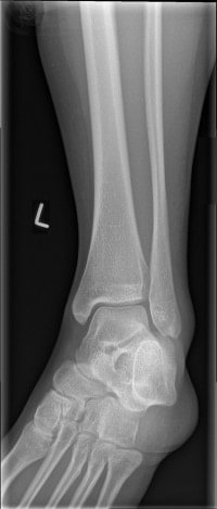 Radiology teaching during medical school is variable, ranging from informal teaching to required clerkships [1]. Many of us likely received an approach to a chest x-ray, but approaches to other studies may or may not have not been taught. We can do better! Enter EM:Rad, a series aimed at providing “just in time” approaches to commonly ordered radiology studies in the emergency department. When applicable, it will provide pertinent measurements specific to management, and offer a framework for when to get an additional view, if appropriate. We recently covered the


 Two patients present to your emergency department: Patient 1 is a 17 year-old soccer player who fell during a game onto their right side and is now complaining of mild right shoulder pain. You obtain x-rays (Figure 1). Patient 2 is a 21 year-old motorist who lost control and went over the handlebars. They heard a pop and are complaining of left shoulder pain. You obtain shoulder x-rays (Figure 2). For these cases, what are your diagnoses, expected physical examination findings, and emergency department management?