ACMT Toxicology Visual Pearl: Sink or Swim

What type of mothball floats in both water and saturated salt solution?
- Camphor
- Naphthalene
- Paradichlorobenzene
- Sodium Hypochlorite



What type of mothball floats in both water and saturated salt solution?

Caring for a patient that is critically-ill secondary to a toxic ingestion is complicated and, in severe cases, extracorporeal treatments (ECTRs) may be considered. The most commonly used ECTRs are intermittent hemodialysis (iHD) and continuous renal replacement therapy (CRRT), but ECTRs also include exchange transfusion, hemoperfusion, liver dialysis, and therapeutic plasma exchange. Finding and evaluating the supporting literature for these treatment modalities in a timely manner is not feasible in most situations. In order to assist in this effort, the EXtracorporeal Treatments In Poisoning (EXTRIP) workgroup has reviewed and provided free, evidence-based recommendations regarding the use of ECTRs for many common toxins and toxicants [1]. These recommendations can be found in a summarized format on the EXTRIP website and the links to their comprehensive reviews are published on PubMed with direct links on their website. This international workgroup is made up of experts in toxicology, nephrology, emergency medicine, pediatrics, pharmacology, critical care, and more. An excellent example of this resource is their review and recommendations on ECTRs for poisoning secondary to beta-adrenergic antagonists (BAAs).
The EXTRIP workgroup included 76 publications in this comprehensive review on the use of ECTRs in BAA poisoning [2]. They evaluated pharmacokinetic/toxicokinetic data for a total of 334 patients poisoned with various BAAs, of which ~90% of the data was published prior to 1990 and does not necessarily represent the improved clearance of these medications with modern ECTR modalities. Based on this evidence, they deemed atenolol, nadolol, and sotalol as dialyzable BAAs. They also reviewed case reports/series of 37 patients with BAA toxicity and made recommendations for those agents with sufficient evidence. Based on the above data, the EXTRIP group recommends iHD over CRRT in patients severely poisoned with atenolol or sotalol and kidney impairment. They make no recommendation for or against ECTR in patients severely poisoned with atenolol or sotalol with normal kidney function and they recommend against ECTR in patients severely poisoned with propranolol.
Read other articles in the EM Pharm Pearls Series and find previous pearls on the PharmERToxguy site.
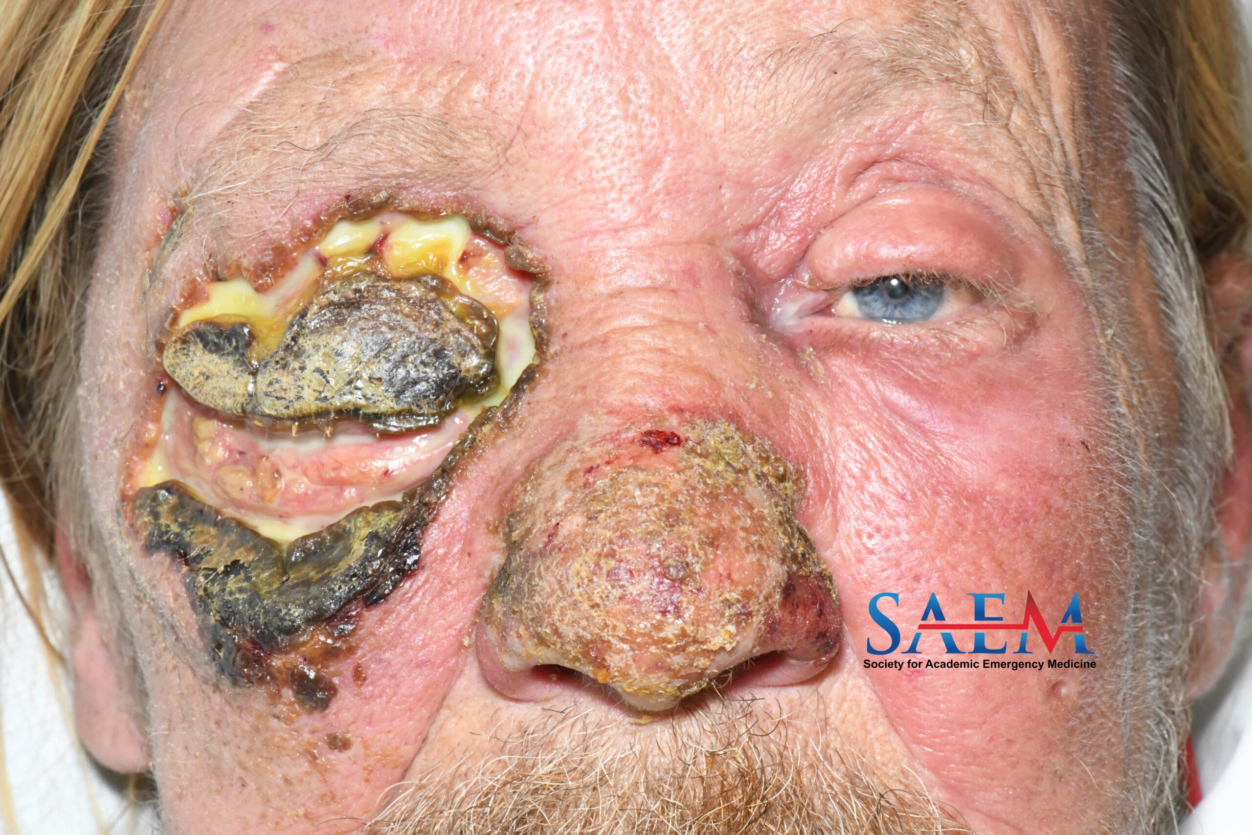
A 59-year-old gentleman experiencing homelessness with a history of hepatocellular carcinoma, hepatitis C, alcohol use disorder, and tobacco dependence presented to the emergency department (ED) with severe, worsening right eye pain, blurry vision, swelling, redness, and purulent discharge after scraping his upper face on concrete during a mechanical fall two weeks prior. Of note, his partner presented to the ED at the same time with a necrotic infection of the breast as well as multiple skin lesions reportedly due to insect bites.
Human trafficking is a devastating crime, where a human being’s labor is exploited through force, fraud, or coercion, for someone else’s profit (1). For survivors, connecting to support in the community can be incredibly difficult, and may come at the expense of their personal safety (1, 2).
The emergency department (ED) is a rare exception, with some studies estimating that over 60% of trafficked persons will present at some point during their exploitation to the ED (3). Unfortunately, less than 5% of emergency physicians report feeling confident in their ability to identify a trafficked person, citing confusion around patient characteristics and their role as a provider (4).
By learning more about human trafficking, ED providers can better prepare themselves to identify and provide appropriate support to those who experience human trafficking.
What can I do to be ready in the ED?
Click to view full-size image. |
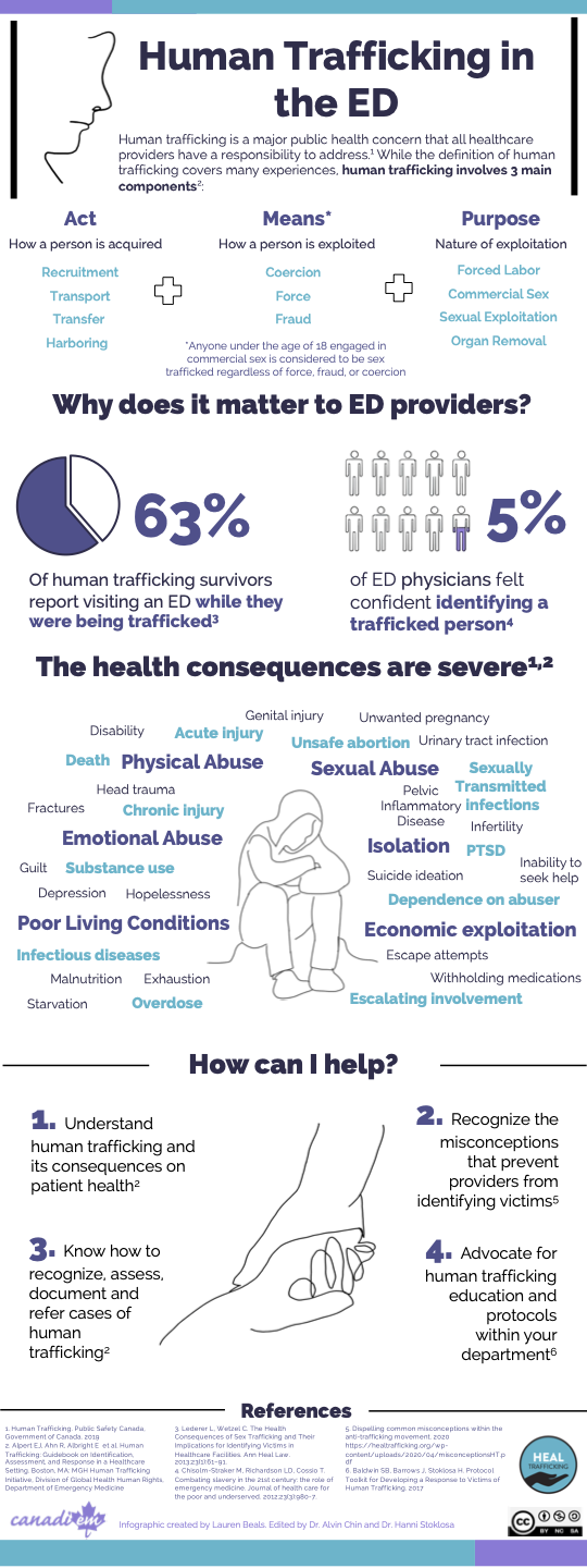 |
Human trafficking always involves 3 components –an act, a means, and a purpose.
Often human trafficking will overlap with other crimes such as assault, domestic violence, rape, and child abuse (5). Of note, anyone under the age of 18 engaged in commercial sex is considered to be sex trafficked regardless of whether a means is present, as they cannot provide consent.
Human trafficking is widespread, but is often undetected, making true estimates of size difficult.
For example, human trafficking prevalence estimates may fail to account for survivors who do not recognize they are being exploited or are afraid to disclose (6, 7).
While no identity is spared, there are certain populations that are at greater risk. These may include:
The only thing all trafficked persons have in common is their vulnerability (1). Trafficking determinants can be conceptualized as “push” and “pull” factors. Push factors lead people to away from their current situation to trafficking (e.g., abuse, poverty, family conflict). Pull factors, drive an individual to something new that increases the risk of trafficking (e.g., income, housing, access to substances) (9, 10).
In the same way that anyone can be trafficked, anyone can be a trafficker.
Traffickers may be well known in the community, recruiting victims from places of employment or education (1). They may be a family member. They may also lure at-risk individuals by acting as a romantic partner, or by providing emotional affirmation, financial assistance, and material goods (1).
A number of tactics can be employed by traffickers, each tailored to the individual survivor but can include any combination of the following (1).
Specific situations to be wary of:
Click to view full-size images
The goal of the visit is NOT to elicit a disclosure.
Your role as a provider is not to investigate or confirm the presence of trafficking, but to respect the autonomy of the patient in front of you, meet their healthcare needs, and empower them to seek additional support on their terms.


Click for full-size images
| Patient Indicators | Companion Indicators |
|---|---|
| Delayed presentation | Refuses to leave |
| Discrepancy between history and clinical presentation | Insists on translating or speaking for the patient |
| Scripted/memorized history | Controlling, interrupting |
| Hypervigilance, fearful | Has patient’s documents in their possession |
| Cannot produce identification | Employer demanding access to medical information |
| Work-related injury with unsafe conditions | |
| Fearful attachment to a cell phone (often used for communication and tracking) |
Any next steps should always be determined by the patient
Unless local criteria for mandatory reporting are met, Police should only be contacted at the explicit instruction of the patient (6, 16).
Consider implementing an ED and institutional protocol for human trafficking. A complete protocol guide is available through HEAL Trafficking.
Planning to write the next great FOAM post on hyperkalemia, but not sure what’s already been done? Or maybe a less popular topic, that you hope has minimal coverage? Or are you a resident, medical student, or even a curious attending searching for the latest, best FOAM regarding a specific topic?
If so, then we have great news for these answers are here in the recently published second Systematic Online Academic Resource (SOAR) Topic Review.1 This series aims to identify and evaluate online education resources by topic. The inaugural entry has already revealed the FOAM landscape related to the renal and genitourinary organ system.2 We can now share results for endocrine, metabolic and nutritional disorders.
We developed search terms specific to endocrine, metabolic and nutritional Disorders from the American Board of Emergency Medicine’s Model of the Clinical Practice of Emergency Medicine (MPCEM). We then performed a systematic search of both FOAMSearch as well as each site within the social media index top 50.3,4 We then assessed the quality of those resources using the revised METRIQ score and designated learner levels for each resource.5
The search yielded 36,346 resources, of which 756 met the criteria for quality assessment. Table 1 displays the 121 posts identified as high quality (rMETRIQ ≥ 16). Overall, the most covered subtopic was potassium disorders, representing 15% of all posts. Subtopics that did not have a high-quality resource identified include metabolic alkalosis, respiratory alkalosis, fluid overload, phosphorus metabolism, hyperglycemia, malabsorption, malnutrition and thyroiditis. Table 2 graphically describes the number of high-quality posts by subtopic coverage and recommended target audience. Of note, resources that were better categorized under a different MCPEM category were generally not included. For example, fluid overload content specific to heart failure was not included.
We hope that educators and learners can use this information to find relevant educational resources, and that FOAM content creators can use this information to help identify topics to produce new content.
| Subtopic | Name of First Author | Name of Blog Post | Level of Trainee Recommendation |
| Metabolic Acidosis | Farkas, Josh | PulmCrit: pH-guided fluid resuscitation & BICAR-ICU | Intern, Junior, Senior, Attending |
| Metabolic Acidosis | Farkas, Josh | Is correcting hyperchloremic acidosis beneficial? | Intern, Junior, Senior, Attending |
| Metabolic Acidosis | Rusyniak, Dan | Tox & Hound – aka AKA | Intern, Junior, Senior, Attending |
| Metabolic Acidosis | Multiple | Alcoholic ketoacidosis – WikEM | Clerk, Intern, Junior, Senior |
| Metabolic Acidosis | Spiegel, Rory | CC Nerd-The Case of the Neutral Documents | Clerk, Intern, Junior, Senior, Attending |
| Respiratory Acidosis | Chan, Wendy | VBG vs ABG in Hypercarbia | Clerk, Intern, Junior, Senior |
| Acid Base (Mixed) | Farkas, Josh | Fluid selection using pH-guided resuscitation | Intern, Junior, Senior, Attending |
| Acid Base (Mixed) | Weingart, Scott | Acid Base in the Critically Ill – Part V – Enough with the Bicarb Already | Intern, Junior, Senior, Attending |
| Corticoadrenal Insufficiency | Long, Brit | Mimics of Sepsis: What do ED Physicians Need to Know? | Intern, Junior, Senior |
| Corticoadrenal Insufficiency | Farkas, Josh | Adrenal crisis – EMCrit Project | Intern, Junior, Senior, Attending |
| Corticoadrenal Insufficiency | Gaillard, Frank | Adrenal hemorrhage | Radiology Reference Article | Radiopaedia.org | Intern, Junior, Senior, Attending |
| Cushing’s Syndrome | Multiple | Cushing syndrome | Radiology Reference Article | Radiopaedia.org | Clerk, Intern, Junior, Senior, Attending |
| Calcium | Long, Brit | Oncologic Emergencies Part I: Pearls and Pitfalls | Intern, Junior, Senior, Attending |
| Calcium | Swaminathan, Anand | Hypercalcemia | Intern, Junior, Senior, Attending |
| Calcium | Multiple | Hypocalcemia – WikEM | Intern, Junior, Senior, Attending |
| Calcium | Taliaferro, Dustin | EM@3AM: Hyper- and HypoCa | Intern, Junior, Senior, Attending |
| Hypovolemia | Astin, Matt | The SPLIT Trial: Saline vs Plasma-Lyte Fluid Therapy | Intern, Junior, Senior, Attending |
| Hypovolemia | Rezaie, Salim | SMART Trial Part 2: Secondary Analysis of Balanced Crystalloids vs Saline in Sepsis | Clerk, Intern, Junior, Senior |
| Hypovolemia | Rezaie, Salim | Is the Great Debate Between Balanced vs Unbalanced Crystalloids Finally Over? | Clerk, Intern, Junior, Senior |
| Hypovolemia | Rezaie, Salim | Does Lactated Ringers (LR) Raise Serum Lactate? | Clerk, Intern, Junior, Senior |
| Hypovolemia | Farkas, Josh | PulmCrit- Get SMART: Nine reasons to quit using normal saline for … | Intern, Junior, Senior, Attending |
| Hypovolemia | Farkas, Josh | IBCC chapter & cast – Fluid selection & pH-guided fluid resuscitation | Intern, Junior, Senior, Attending |
| Hypovolemia | Farkas, Josh | The SPLIT trial: Internal vs. external validity | Intern, Junior, Senior, Attending |
| Hypovolemia | Rezaie, Salim | Balanced vs Unbalanced Fluids in Pediatric Severe Sepsis | Clerk, Intern, Junior, Senior |
| Hypovolemia | Multiple | Fluid Choice in Sepsis: Does it matter? | Clerk, Intern, Junior, Senior, Attending |
| Hypovolemia | Kenny, Jon-Emile | Are balanced crystalloids better than saline? SMART Talk with Dr … | Intern, Junior, Senior, Attending |
| Hypovolemia | Farkas, Josh | PulmCrit- Overcoming occult diuretic resistance: Achieving diuresis … | Attending |
| Hypovolemia | Multiple | CT hypoperfusion complex | Radiology Reference Article … | Clerk, Intern, Junior, Senior, Attending |
| Hypovolemia | Multiple | Dehydration (peds) – WikEM | Clerk, Intern, Junior, Senior, Attending |
| Hypovolemia | Rezaie, Salim | The Great Debate Between Balanced and Unbalanced Crystalloids Continues | Junior, Senior, Attending |
| Hypovolemia | Morgenstern, Justin | IV fluid choice part 1: The SPLIT trial | Clerk, Intern, Junior, Senior, Attending |
| Hypovolemia | Drenzla, Adam | SALT OF THE EARTH (PART 1) | Clerk, Intern, Junior, Senior |
| Potassium | Meyers, Pendell | Critical Hyperkalemia by H. Pendell Meyers, EMCrit Intern – EMCrit | Junior, Senior, Attending |
| Potassium | Farkas, Josh | Hyperkalemia – EMCrit Project | Clerk, Intern, Junior, Senior |
| Potassium | Farkas, Josh | Management of severe hyperkalemia in the post-Kayexalate era | Intern, Junior, Senior, Attending |
| Potassium | Swaminathan, Anand | Is Kayexalate Useful in the Treatment of Hyperkalemia in the … | Clerk, Intern, Junior, Senior |
| Potassium | Singh, Manpreet | Hyperkalemia – The Great Imitator | Clerk, Intern, Junior, Senior |
| Potassium | Multiple | Tumor Lysis Syndrome – Diagnosis and Treatment | Clerk, Intern, Junior |
| Potassium | Multiple | Management of the Sick Dialysis/ESRD Patient | clerk, intern, Junior, senior |
| Potassium | Meyers, Pendell | A young man with back spasms | Intern, Junior, Senior |
| Potassium | Helman, Anton | Emergency Management of Hyperkalemia | EM Cases Podcast | Junior, Senior, Attending |
| Potassium | Long, Brit | Updates in Management of Hyperkalemia | Clerk, Intern, Junior, Senior |
| Potassium | Weingart, Scott | Treatment of hyperkalemia in the ED | Clerk, Intern, Junior, Senior |
| Potassium | Farkas, Josh | PulmCrit- BRASH syndrome: Bradycardia, Renal failure, Av blocker … | intern, Junior, senior, attending |
| Potassium | Farkas, Josh | Myth-busting: Lactated Ringers is safe in hyperkalemia, and is … | Junior, Senior, Attending |
| Potassium | Multiple | Insulin Dosing in Hyperkalemia | Intern, Junior, Senior, Attending |
| Potassium | Rezaie, Salim | ECG Changes of Hyperkalemia | Clerk, Intern, Junior, Senior |
| Potassium | Santistevan, Jamie | emDOCs.net – Emergency Medicine EducationSubtle ECG findings … | Clerk, Intern, Junior, Senior, Attending |
| Potassium | Morton, A. Ross | Management of Hyperkalemia with ECG Changes | clerk, intern, Junior, senior |
| Potassium | Awad, Nadia | Thyroid Storm: Treatment Strategies | Intern, Junior, Senior, Attending |
| Potassium | Grock, Andrew | Bicarbonate: Completely Useless? | Junior, Senior, Attending |
| Potassium | Hayes, Bryan D | Hyperkalemia Management: Preventing Hypoglycemia From Insulin | Senior, Attending |
| Sodium | Smarandache, Andrei | When and how to treat hyponatremia in the ED | Intern, Junior, Senior |
| Sodium | Farkas, Josh | Hypernatremia & dehydration in the ICU – EMCrit Project | Senior, Attending |
| Sodium | Farkas, Josh | Emergent treatment of hyponatremia or elevated ICP with bicarb … | intern, Junior, senior, attending |
| Sodium | Farkas, Josh | Taking control of severe hyponatremia with DDAVP | Attending |
| Sodium | Helman, Anton | Emergency Management of Hyponatremia | EM Cases | intern, Junior, senior, attending |
| Sodium | Farkas, Josh | Unconventional therapies for hyponatremia: Thinking outside the … | intern, Junior, senior, attending |
| Sodium | Farkas, Josh | PulmCrit- Controlled aquaresis: Management of hypervolemic or euvolemic hyponatremia with oral urea | Junior, senior, attending |
| Magnesium | Morgenstern, Justin | Torsades de Pointes: Approach to resuscitation | intern, Junior, senior |
| Magnesium | Multiple | Hypermagnesemia – WikEM | Clerk, Intern, Junior, Senior, Attending |
| Diabetic Ketoacidosis | Lodeserto, Frank | Pediatric DKA: Do Fluids Really Matter? – REBEL EM – Emergency … | Clerk, Intern, Junior, Senior |
| Diabetic Ketoacidosis | Morgenstern, Justin | IV fluids do not cause cerebral edema in pediatric DKA – First10EM | Clerk, Intern, Junior, Senior, Attending |
| Diabetic Ketoacidosis | Farkas, Josh | Blood gas measurements in DKA: Are we searching for a unicorn? | Intern, Junior, Senior, Attending |
| Diabetic Ketoacidosis | Farkas, Josh | Anatomy of a DKA resuscitation – EMCrit Project | Clerk, Intern, Junior, Senior |
| Diabetic Ketoacidosis | Hughes, Darrel | Is There Any Benefit to an Initial Insulin Bolus in Diabetic … | Clerk, Intern, Junior, Senior, Attending |
| Diabetic Ketoacidosis | Abela, Nikki | Fluid Type and Infusion Rate in Paediatric DKA | Clerk, Intern, Junior, Senior, Attending |
| Diabetic Ketoacidosis | Multiple | Cerebral edema in DKA – WikEM | Clerk, Intern, Junior, Senior, Attending |
| Diabetic Ketoacidosis | Lubberdink, Ashley | TREKK Series | Diabetic Ketoacidosis | Clerk, Intern, Junior |
| Diabetic Ketoacidosis | Farkas, Josh | PulmCrit- Dominating the acidosis in DKA | Intern, Junior, Senior, Attending |
| Diabetic Ketoacidosis | Farkas, Josh | PulmCrit – Four DKA Pearls | Intern, Junior, Senior, Attending |
| Diabetic Ketoacidosis | Multiple | emDOCs.net – Emergency Medicine Education Diabetic … | Clerk, Intern, Junior, Senior, Attending |
| Diabetic Ketoacidosis | Multiple | Diabetic ketoacidosis | Radiology Reference Article | Radiopaedia.org | Clerk, Intern, Junior, Senior, Attending |
| Diabetic Ketoacidosis | Multiple | PEM Pearls: Treatment of Pediatric Diabetic Ketoacidosis and the Two-Bag Method | Junior, Senior, Attending |
| Diabetic Ketoacidosis | Monette, Derek | PEM Practice Changing Paper: Clinical Trial of Fluid Infusion Rates for Pediatric DKA | Junior, Senior, Attending |
| Hyperosmolar Hyperglycemic State | Multiple | Hyperosmolar hyperglycemic state – WikEM | Clerk, Intern, Junior, Senior, Attending |
| Hyperosmolar Hyperglycemic State | Multiple | Hyperosmolar hyperglycemic state | Radiology Reference Article … | Clerk, Intern, Junior, Senior, Attending |
| Hyperosmolar Hyperglycemic State | Multiple | Non-ketotic hyperglycaemic seizure | Radiology Reference Article … | Clerk, Intern, Junior, Senior, Attending |
| Hypoglycemia | Multiple | Hypoglycemic encephalopathy | Radiology Reference Article … | Clerk, Intern, Junior, Senior, Attending |
| Insulin Pump | Multiple | Insulin Pumps: Complications and Emergency Department Presentations | Intern, Junior, Senior, Attending |
| Insulin Pump | Weingart, Scott | Podcast 198 – Insulin Pumps and Such with Josh Miller, MD | Clerk, Intern, Junior, Senior, Attending |
| Insulin Pump | Rezaie, Salim | Diabetic Gastroparesis Needs HUGS | Intern, Junior, Senior, Attending |
| Nutritional Disorders | Multiple | Osteoporosis | Radiology Reference Article | Radiopaedia.org | Clerk, Intern, Junior, Senior, Attending |
| Nutritional Disorders | Long, Brit | Protein Shakes and Dietary Supplements: What are their ingredients and how much is too much? | Senior, Attending |
| Vitamin Deficiencies | Multiple | Rickets | Radiology Reference Article | Radiopaedia.org | Clerk, Intern, Junior, Senior, Attending |
| Vitamin Deficiencies | Groth, Meghan | Mythbusting the Banana Bag | Intern, Junior, Senior, Attending |
| Wernicke-Korsakoff | Multiple | Thiamine Deficiency | Intern, Junior, Senior, Attending |
| Wernicke-Korsakoff | Pescatore, Rick | Should You Prescribe Oral Thiamine for Chronic Alcoholics … | Senior, Attending |
| Wernicke-Korsakoff | Multiple | Wernicke-Korsakoff syndrome – WikEM | Clerk, Intern, Junior, Senior, Attending |
| Wernicke-Korsakoff | Multiple | Wernicke encephalopathy | Radiology Reference Article … | Clerk, Intern, Junior, Senior, Attending |
| Wernicke-Korsakoff | Swaminathan, Anand | Wernicke’s Encephalopathy Archives – REBEL EM – Emergency … | Junior, Senior, Attending |
| Parathyroid Disease | Multiple | Parathyroid hormone | Radiology Reference Article | Radiopaedia.org | Clerk, Intern, Junior, Senior, Attending |
| Parathyroid Disease | Multiple | Parathyroid glands | Radiology Reference Article | Radiopaedia.org | Clerk, Intern, Junior, Senior, Attending |
| Parathyroid Disease | Newman, Samantha | PTH versus PTHrP — Small Differences, Big Implications – Clinical … | Intern, Junior, Senior |
| Pituitary Disorders | Multiple | Sheehan syndrome | Radiology Reference Article | Radiopaedia.org | Clerk, Intern, Junior, Senior, Attending |
| Pituitary Disorders | Multiple | emDOCs.net – Emergency Medicine EducationThunderclap … | Intern, Junior, Senior |
| Pituitary Disorders | Multiple | Pituitary gland | Radiology Reference Article | Radiopaedia.org | Clerk, Intern, Junior, Senior, Attending |
| Pituitary Disorders | Multiple | Optic pathway glioma | Radiology Reference Article | Radiopaedia.org | Clerk, Intern, Junior, Senior, Attending |
| Pituitary Disorders | Multiple | Pituitary apoplexy | Radiology Reference Article | Radiopaedia.org | Intern, Junior, Senior, Attending |
| Pituitary Disorders | Multiple | Empty sella | Radiology Reference Article | Radiopaedia.org | Intern, Junior, Senior, Attending |
| Pituitary Disorders | Multiple | Elevated prolactin (differential) | Radiology Reference Article … | Clerk, Intern, Junior, Senior, Attending |
| Hyperthyroid | Multiple | Thyroid storm – WikEM | Intern, Junior, Senior, Attending |
| Hyperthyroid | Lipp, Chris | CRACKCast E128 – Thyroid and Adrenal Disorders | Intern, Junior, Senior, Attending |
| Hyperthyroid | Multiple | Hyperthyroidism – WikEM | Clerk, Intern, Junior, Senior |
| Hyperthyroid | Multiple | Thyroid Storm and Aortoiliac Occlusive Disease | Intern, Junior, Senior, Attending |
| Hyperthyroid | Multiple | Bedside Rounds Series: Goiter – Clinical Correlations | Clerk, Intern, Junior, Senior |
| Hyperthyroid | Lee, Terrance | Diagnosing hyperthyroidism: Answers to 7 common questions | Clerk, Intern, Junior, Senior |
| Hypothyroid | Fox, Sean | Hypothyroidism and Myxedema Coma — Pediatric EM Morsels | Intern, Junior, Senior, Attending |
| Hypothyroid | Multiple | Myxedema coma – WikEM | Intern, Junior, Senior, Attending |
| Hypothyroid | Zhao, Leah | Spot the Diagnosis! The case of the Pale Woman | Intern, Junior, Senior |
| Tumors Of Endocrine Glands | Multiple | Parathyroid adenoma | Radiology Reference Article | Radiopaedia.org | Clerk, Intern, Junior, Senior, Attending |
| Tumors Of Endocrine Glands | Multiple | Central nervous system germinoma | Radiology Reference Article … | Intern, Junior, Senior, Attending |
| Tumors Of Endocrine Glands | Multiple | Craniopharyngioma | Radiology Reference Article | Radiopaedia.org | Junior, Senior, Attending |
| Tumors Of Endocrine Glands | Multiple | Carcinoid tumor | Radiology Reference Article | Radiopaedia.org | Intern, Junior, Senior, Attending |
| Adrenal Tumor | Multiple | Pheochromocytoma | Radiology Reference Article | Radiopaedia.org | Intern, Junior, Senior, Attending |
| Adrenal Tumor | Multiple | Adrenal adenoma | Radiology Reference Article | Radiopaedia.org | Intern, Junior, Senior, Attending |
| Adrenal Tumor | Multiple | Adrenal cortical carcinoma | Radiology Reference Article … | Intern, Junior, Senior, Attending |
| Pituitary Tumor | Multiple | Acromegaly | Radiology Reference Article | Radiopaedia.org | Clerk, Intern, Junior, Senior, Attending |
| Pituitary Tumor | Multiple | Pituitary adenoma | Radiology Reference Article | Radiopaedia.org | Clerk, Intern, Junior, Senior, Attending |
| Thyroid Tumor | Multiple | Medullary thyroid carcinoma | Radiology Reference Article … | Clerk, Intern, Junior, Senior |
| Thyroid Tumor | Multiple | Anaplastic thyroid carcinoma | Radiology Reference Article … | Intern, Junior, Senior, Attending |
References:
Special thanks to SOAR co-authors Ryan Pedigo, Lisa Zhao, JooYeon Jung, Shirley W Bae, Teresa Chan, and Andrew Grock.
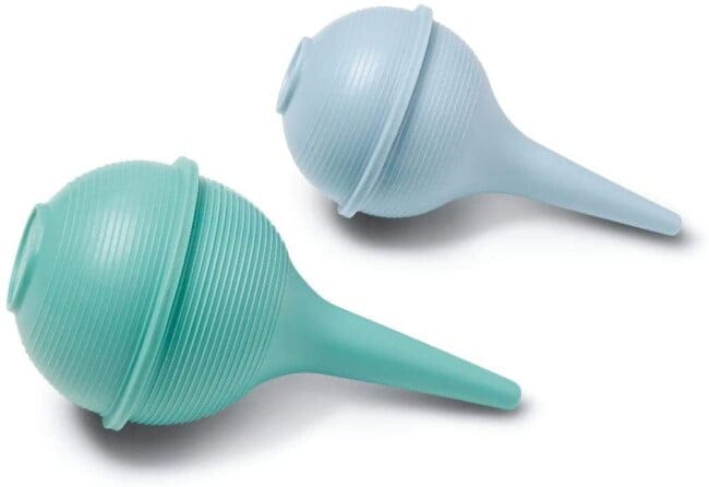
Nasal congestion is a common symptom of upper respiratory tract infections, such as bronchiolitis, in newborns and infants. Because newborns are obligate nose breathers, any congestion presents a challenge during feeding and sleeping. These infants become frustrated when they cannot breathe while feeding and tend to have disturbed sleep when their nasal passages are occluded. This often leads to dehydration and irritability. Although the infant bulb syringe (above) can often alleviate the congestion, other commercial products may be able to more forcefully clean out the nasal mucus (e.g., NoseFrida, Bubzi Nasal Aspirator).
In the Emergency Department, you may encounter families who may not have the resources to purchase or be aware of commercial aspiration devices for children. The concept behind our DIY Nasal Snot Aspirator is to allow the caregiver to suction the child’s nose using the negative pressure generated from the caregiver’s own mouth. The left video demonstrates how the NoseFrida works, and the right video demonstrates our DIY Nasal Snot Aspirator. Note that the specimen trap serves as the protective “filter”, or barrier, between the child’s suctioned mucus and the caregiver’s mouth. Thanks to Stephany Landry, RN, BSN for sharing this trick of the trade.
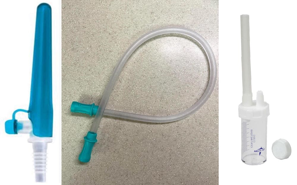
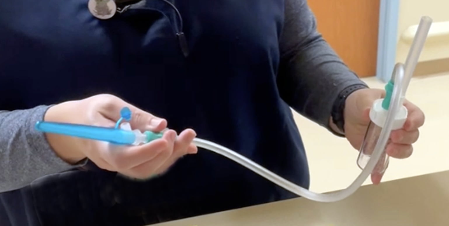
The authors and ALiEM do not have any affiliation with any of these device companies.
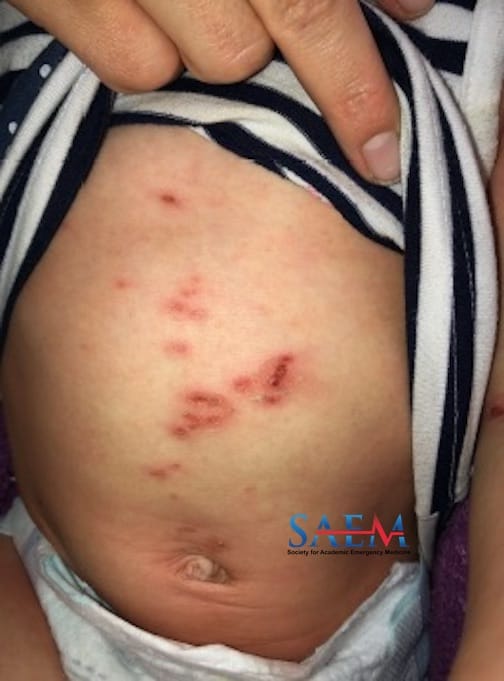
A 17-month-old girl with a history of eczema presents to the pediatric emergency department for evaluation of a rash. The rash is different from her usual eczema, developed three days prior to presentation, and is described as red with yellow crusting. Her mother also noticed blistering in her groin and under her axilla. She has associated fussiness and decreased feeding, but no fever.
Vitals: T 37.7°C; HR 161; BP 115/75; RR 24; O2 sat 100% on room air General: Fussy but consolable Eyes: No conjunctival erythema or discharge Mouth: Yellow crusting and fissuring surrounding mouth; no intra-oral lesions Neck: No nuchal rigidity Cardiovascular: Tachycardic with regular rhythm; no murmurs Respiratory: Normal rate; normal breath sounds Abdomen: Non-tender to palpation; non-distended; normal bowel sounds Neurologic: Alert Skin: Diffusely erythematous; scaling rash over the face with areas of yellow crusting; erythematous areas with blistering/desquamation to the anterior trunk, axilla, and inguinal regions Complete blood count (CBC) and comprehensive metabolic panel (CMP) unremarkable. Staphylococcal scalded skin syndrome caused by impetigo. This case describes a patient with a rash, blistering/desquamation of axilla and groin, and systemic symptoms consistent with staphylococcal scalded skin syndrome (SSSS). Clinical features of SSSS include erythema to intertriginous areas, rapid progression of erythema, and systemic symptoms such as fever, irritability, and poor oral intake. Mucous membranes are not typically involved. Physical exam findings include perioral crusting and fissuring (seen in photo), blanching erythema, desquamation, shallow skin erosions, and a positive Nikolsky sign. SSSS is caused by exfoliative toxin A (ETA) and exfoliative toxin B (ETB), two exotoxins produced by certain strains of S. aureus. ETA and ETB cause the breakdown of keratinocyte adhesions within the epidermis, leading to desquamation. Infection with S. aureus at any site can cause SSSS, including bacterial conjunctivitis, wound/skin infection, staphylococcal pneumonia, pyomyositis, septic arthritis, and endocarditis. SSSS is more common in children, a phenomenon thought to be due to a lack of protective antibodies against staphylococcal antigens. The diagnosis of SSSS is clinical but can be confirmed with histopathology. In this case, the extensive yellow, crusting lesions of the face suggest impetigo, a superficial skin infection predominantly caused by S. aureus, as the etiology of SSSS. Children with eczema are at increased risk of impetigo due to disruption of the normal skin barrier. Complications of SSSS include fluid losses due to extensive skin breakdown, electrolyte abnormalities, sepsis, and death. Penicillinase-resistant penicillins (oxacillin, nafcillin) or first- or second-generation cephalosporins. Clindamycin monotherapy should be avoided due to high rates of resistance. Management of SSSS in most cases also includes hospitalization for IV antibiotics and supportive care. In patients with significant skin involvement, admission to either an ICU or burn unit is warranted for close monitoring and wound care.

Take-Home Points