PEM POCUS Series: Confirmation of Endotracheal Tube Placement

Read this tutorial on the use of point of care ultrasonography (POCUS) for confirmation of endotracheal tube (ETT) placement in pediatric patients. Then test your skills on the ALiEMU course page to receive your PEM POCUS badge worth 2 hours of ALiEMU course credit.
Module Goals
- List indications for performing airway/lung POCUS to confirm ETT placement
- Describe the technique of performing airway and focused lung POCUS
- Distinguish between normal and abnormal airway and lung POCUS findings
- Distinguish between tracheal, endobronchial, and esophageal placement of ETT
- List the limitations of airway and lung POCUS
Case Introduction: The Postictal Toddler
Joey is a 2-year-old male with a history of epilepsy who presents to a community hospital emergency department with generalized tonic-clonic seizures of more than 45 minutes duration. After receiving 2 doses of IV midazolam, he stopped seizing. He has very shallow breathing and oxygen saturations as low as 90 percent on 2 liters of supplemental oxygen via nasal cannula. The pediatric transport team arrives to transport him to another hospital for admission and note that he is somnolent with poor respiratory effort. His current vital signs:
| Vital Sign | Finding |
|---|---|
| Temperature | 37.0 C |
| Heart Rate | 115 bpm |
| Blood Pressure | 85/65 |
| Respiratory Rate | 12 |
| Oxygen Saturation (room air) | 92% on 2 L via nasal cannula |
An end tidal carbon dioxide (ETCO2) monitor shows a ETCO2 level in the high 70s mmHg. The decision is made to intubate the patient given disordered breathing, hypercapnia, and hypoxia following medical management of seizures. The transport team would like to use POCUS to evaluate ETT placement at the outside hospital and during transport.
For simplicity, this module will focus on 3 modes of using POCUS for ETT confirmation. Collectively, these techniques can help improve evaluation.
There are many benefits of using POCUS to confirm ETT placement, such as in the following examples:
- When compared to auscultation, POCUS ETT can be done in a loud environment where auscultation may be challenging (i.e., as may occur in transport or on scene).
- When compared to radiography, POCUS ETT can be done rapidly at the bedside when chest radiography may be delayed or unavailable (i.e., in transport or during chest compressions).
- When compared to capnography, POCUS ETT is helpful in scenarios of low pulmonary blood flow as in cardiac arrest or with poor tissue perfusion when capnography may be less reliable. Also POCUS can distinguish between tracheal and endobronchial ETT placement, whereas capnography cannot.
- Unlike auscultation and capnography, POCUS ETT can confirm placement in real time, even before ventilating the patient, unlike auscultation and capnography to work.
- POCUS ETT should typically be used as an adjunct to other methods of confirmation or in resource-limited settings, if other methods are not available.
Just as all methods of confirming ETT placement have their limitations, so does POCUS. This will be discussed in greater detail later in the module.
There are many factors to consider in the performance of ETT POCUS:
| Factor | Options |
|---|---|
| Probe selection | Linear or curvilinear |
| Location on the anterior neck | Suprasternal notch, cricoid, or thyroid cartilage |
| Probe orientation | Longitudinal or transverse plane |
| Timing | Dynamic (while intubating) or static (for confirmation) |
| Evaluation technique | Direct (visualize the ETT) or indirect (visualize lung movement0 |
Probe Selection
Two types of probes will be needed for POCUS ETT confirmation.
- Use a linear probe to visualize the superficial airway and lung structures. The linear probe uses high frequency sound waves to create high resolution images of superficial structures such as the trachea and pleura.
- Use a curvilinear probe to visualize deeper structures, such as the diaphragm.
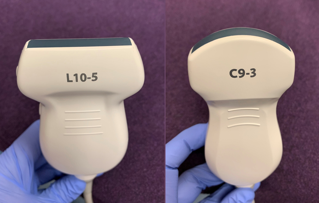
Figure 1: Linear probe (left) and curvilinear probe (right)
Timing of Image Acquisition
If time permits, pre-scan the patient’s neck to locate the trachea. Adjust the gain and depth accordingly to visualize the trachea clearly in the middle of the screen.
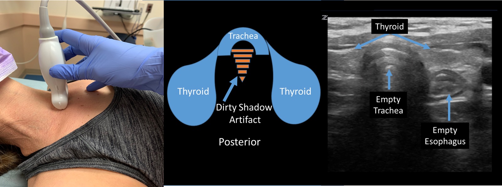
Figure 2: Positioning and ultrasound images of the anterior neck anatomy for ETT placement confirmation. Left: Transverse orientation of the linear probe just above the suprasternal notch. Center: Corresponding pictorial display of the trachea and surrounding structures. Note that below the trachea is a dirty shadow artifact, resulting from the air-mucosa interface. Right: Corresponding ultrasound image of the thyroid lobes flanking the empty trachea, with the ovoid esophagus seen posterolaterally (ultrasound image by Jade Sequin).
1. Static Assessment
- We recommend using the static assessment (i.e., after the patient is intubated), rather than dynamic (i.e., watching the ETT enter the trachea in real time) which is technically more challenging.
- Positioning: Stand at the patient’s waist, facing the patient’s head, with the probe marker pointing towards the patient’s right (transverse plane) to confirm ETT placement in the neck. Place the linear transducer midline on the anterior neck, slightly above the suprasternal notch (figure 2, left). The orientation of the image on the screen corresponds to the probe direction. This orientation is helpful for procedural POCUS and conceptually allows for easier redirection.
- Identify the trachea: The trachea is visible in the midline as a semicircular structure with a hyperechoic bright line (upside down U) and shadows distally (figure 2, center). Shadows are reverberation artifact from the air in the trachea (often called “dirty shadows,” or referred to as the air-mucosa interface). The thyroid overlies the trachea as a homogenous structure with the lobes extending bilaterally.
- Identify the esophagus: The esophagus is generally posterolateral and to the left of the trachea. The esophagus is seen as a collapsed round or oval shaped structure with concentric layers, without air in it (figure 2, right).
- Anatomy variability: A pediatric study noted that the esophagus can be seen in variable locations in relation to the cricoid ring and trachea. It was partially to the patient’s left (62%), completely to the left (20%), behind the cricoid ring (16%), and partially to the right (2%) [1].
When the ETT is placed correctly in the trachea, you should still see only a SINGLE air-mucosa interface, similar to an empty trachea. An ETT properly positioned in the trachea will have a similar ultrasonographic appearance with one air-mucosal interface as the air-filled tube will be in the trachea and the esophagus will be decompressed without air (figure 2, right).
2. Dynamic Assessment
Dynamic assessment involves watching the ETT pass into the trachea in real-time. In this technique, you will see a brief disturbance within the trachea termed the “snowstorm” which is a subtle finding (Video 1). A dynamic assessment is made more challenging with the multiple tasks and personnel at the bedside during intubation.
Video 1: Dynamic assessment of ETT placement confirmation using a linear probe in the transverse orientation on the anterior neck . With the probe marker to the patient’s right, the trachea is often on the left of the screen in relationship to the esophagus, as in this video. As the ETT enters the trachea, there is a slight disruption termed a “snowstorm” noted in this dynamic view. Video credit: Jade Sequin
Erroneous Esophageal Intubation
If the ETT is placed incorrectly in the esophagus, there will be TWO air-mucosa interfaces with reverberation artifact and posterior shadowing. This has been called the “double trachea sign” or “double tract sign” (figure 3, left). Contrast this to normal anatomy with an empty esophagus (figure 3, right).

Figure 3. Left: Double tract or double trachea sign on ultrasound, visualized when the ETT is placed incorrectly in the esophagus. Note the esophagus appears curved with dirty shadow artifact like the trachea. Right: Normal collapsed esophagus. Images credit: Jade Sequin.
Video 2: Esophageal intubation seen on ultrasound. Note the ETT entering the esophagus, generating the “double tract” or “double trachea” sign. Video used with permission by authors of [2].
Video 3: “Double tract” or “double trachea” sign and esophageal de-intubation. The video starts with the ETT in the esophagus, but then is removed. Video used with permission by authors of [2].
This indirect visualization method uses ultrasound to identify bilateral lung sliding as a means to confirm ETT placement, because this implies that both lungs are ventilated. This method is often used in conjunction with and after direct confirmation using POCUS, seeing the ETT in the trachea.
- If the ETT is in the right main stem bronchus, ONLY the right lung will have sliding.
Ultrasound Probe Placement
Place the linear transducer on the superior, most-anterior chest wall in the mid clavicular line over the 3rd-5th intercostal space. Ensure that the probe marker is towards the head. Scan both lungs (Figures 4).
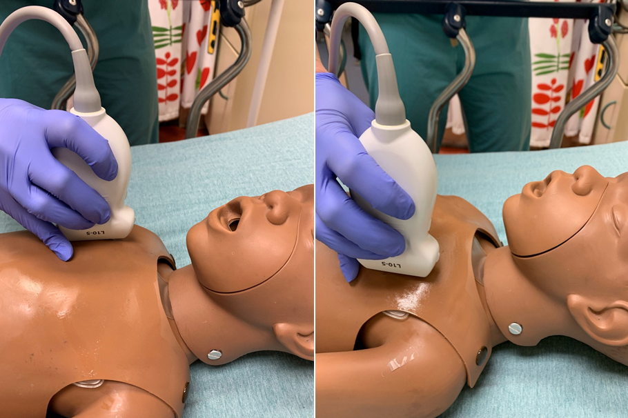
Figure 4. Positioning of the linear probe on the patient’s anterior chest wall to check for lung sliding
Normal Lung Findings on POCUS
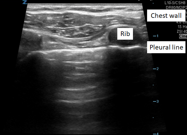
Figure 5. Ultrasound of a normal lung: Just deep to the chest wall and ribs, the pleural line of the lung slides horizontally to and fro with each breath.This line is the first hyperechoic line deep to the rib and is the place to look for lung sliding.
Alveoli filled with air have the ARTIFACTS that are the hallmark of airway POCUS.
- A lines (figure 6): Hyperechoic lines that are parallel to the pleural line (typically horizontal) that are caused by reverberations between the pleura and transducer. They are equidistant from the chest wall. A lines are seen with normal aerated lungs along with lung sliding
- Z lines or comet tails: Perpendicular lines to the pleura (often appear vertical as the pleura is typically visualized as horizontal) that arise from the pleura. These lines typically do not go to the bottom of the screen.
- Lung sliding (figure 8): Shimmering artifact of the parietal and visceral pleura sliding against each other. Lung sliding indicates that the lung visualized under the probe is filled with air and ventilated (video 4).

Figure 6. Normal lung with A lines – The most superficial hyperechoic line below the chest wall is the pleural line. The subsequent hyperechoic lines parallel and deep to the pleural line are A lines. A lines are always normal findings.
Video 4: Normal lung ultrasound: Most superficial are the chest wall tissue and 2 ribs (the circular anechoic structures). The hyperechoic line just deep to the ribs is the pleural line. Lung sliding is the subtle movement at the pleural line, referred to as “ants marching.” The hyperechoic lines horizontal and parallel to the pleural line are A lines, and the thin vertical lines are Z lines, or comet tails.
B Lines
In contrast to A lines, B lines may be visualized in patients with abnormal lungs. B lines are hyperechoic lines (typically vertical) that arise at the pleural line and go all the way to the bottom of the screen (at least 4-8 cm depth with some experts recommending to 16 cm). This is in contrast to Z lines which do not go to the bottom of the screen. The presence of multiple B lines indicates increased fluid in the interstitium of the lungs, which can be seen in conditions such as bronchiolitis and pulmonary edema (figure 7, videos 5 and 6). Note that the presence of B lines also indicate aerated lungs.

Figure 7. Lung POCUS showing A and B lines. A lines are the hyperechoic lines parallel to the pleural line. B lines are the hyperechoic projections perpendicular to the pleural line that extends to the bottom of the screen. A lines are normal, while multiple B lines may be pathogenic.
Video 5: Lung ultrasound showing multiple hyperechoic, perpendicular B lines.
Video 6: Lung ultrasound showing lung sliding and multiple B lines. Note that this image uses a curvilinear probe.
M-Mode Setting
For additional confirmation of lung sliding, press the M mode button (motion mode) without lifting the probe to visualize motion of the sliding pleura. The M-mode view represents a small narrow slice of the ultrasound image (where the bold white vertical line appears) and runs only that portion over time.
- Lung is aerated: Looking below the pleural line level ,you will see a grainy display, known as the “sandy beach” or “seashore” signs (figure 8). You’ll find yourself feeling very relaxed when you see this, because this indicates a successfully aerated lung.
- Lung is NOT aerated: Looking below the pleural line level, you will see multiple horizontal bar-like, striated lines instead of the grainy, sandy beach (figure 9). This is called the “barcode” or “stratosphere” sign, and may be seen in a pneumothorax or a main-stem bronchus intubation.

Figure 8: Lung ultrasound with M-mode view in a normal, aerated lung (left), showing the grainy, “sandy beach” appearance of the lines deep to the pleural line. Contrast this to an abnormal, non-aerated lung (right), showing the horizontal “barcode” appearance of the lines deep to the pleural line.

Figure 9: Another example of a normal (left) and non-aerated (right lung) in M-mode view
Ultrasound Technique
Visualize lung sliding in both 2D (also known as B mode and is the typical ultrasound mode) and M mode on the both the left and right chest.
- Note: If the ETT is in the right mainstem bronchus, you may still see subtle movements of the pleural line on the left due to cardiac activity. The lung sliding in this case will be asymmetric with less movement of the pleural line on the left compared to right.
Alternative Causes for Abnormal Lung Sliding After Intubation
Abnormal lung sliding on ultrasound may be worrisome for an esophageal intubation, because the lungs are not aerated with PPV breaths. However, there are other causes to consider before removing the ETT for a re-intubation attempt.
1. Pneumothorax
In order to see lung sliding, visceral and parietal pleural need to be touching. With a pneumothorax, there is air in the pleural space. The parietal pleura will still be visible, but the visceral pleura and moving interface are not seen. In the M-mode view, a “barcode sign” will be present (figure 10), highlighting the importance of evaluating both 2D (B mode) and M mode if there is any doubt about lung sliding.

Figure 10: Lung POCUS demonstrating no lung sliding (“barcode sign”) in M-mode view
Video 10: Lung POCUS of a patient with a pneumothorax, showing no lung sliding for one lung in 2D view (B mode)
2. Main stem bronchus intubation
If there is no lung sliding in just one lung (especially if it occurs on the left), this may be caused by the ETT being too deep into a mainstem bronchus. This results in non-ventilation of the contralateral lung. Be aware that since the visceral and parietal pleural are still touching (unless there is also a pneumothorax), you could see some sliding movement, as the heart still causes some movement of the lungs.
3. ETT obstruction or apnea
This results in the loss of lung sliding bilaterally.
Take Away
When you see symmetric lung sliding on both sides of the chest, the ETT is in good position in the trachea.
Ultrasound Probe Placement
Use a curvilinear probe, because it gives you deeper tissue penetration than the linear probe. This allows you to better visualize the diaphragm, which is a deeper structure.

Figure 11. Left: Using a curvilinear probe with the probe marker towards the head, position it along the mid-axillary line to identify the diaphragm. Continue sliding the probe to the lower edge of the ribcage until you see the diaphragm meeting the spine along the bottom of the ultrasound image. Right: Ideal ultrasound view of the hyperechoic diaphragm. Also seen is the liver with mixed echotexture, a hypoechoic kidney, and the hyperechoic spine.
Normal Findings on POCUS (figure 11)
- The diaphragm is a hyperechoic line, seen curving vertically on the screen, with a solid organ (liver or spleen) caudal to that.
- The spine appears as interrupted hyperechoic structures (vertebral bodies), extending caudally from the diaphragm at the bottom of the image. The vertebral bodies shadow as all calcified structures on ultrasound do. Normally the spine is only visualized caudal to the diaphragm, because aerated lung obscures visualizing the spine in the thorax (cephalad to the diaphragm).
Ultrasound Technique
- Watch the movement of the diaphragm. In a patient who is paralyzed for intubation, the diaphragm will only move with delivery of positive pressure ventilation (PPV).
- Normal: If the ETT is in good position, with a PPV breath, the diaphragm moves caudal toward the abdomen as the lungs inflate, and upwards when the lungs deflate (video 7). In M mode, normal diaphragm movement creates a smooth wave with inspiration and expiration (video 8).
- Esophageal intubation: The diaphragm moves in the reverse direction than is expected. With a PPV breath, the diaphragm moves cephalad, because the abdominal cavity is getting inflated.
- Mainstem bronchus intubation: The diaphragm on the side of the main stem intubation (typically right) will show exaggerated motion toward the abdomen during PPV. The diaphragm on the contralateral side, where the lung is not properly ventilated will either not move or move paradoxically cephalad during PPV. In M-mode, there is no sinusoidal, wave pattern for the diaphragm in the non-ventilated lung (video 9)
Video 7: Ultrasound view showing diaphragmatic movement with regular breaths. The diaphragm pushes the spleen and kidneys caudal into the abdomen (to the right of the screen) with each breath.
Video 8: Ultrasound M-mode view of the diaphragm with regular breaths. Normal diaphragmatic movement is demonstrated by the hyperechoic sinusoidal line (at 12 cm depth) at the bottom of the screen.
Video 9: Ultrasound of the diaphragm in M-mode setting. The hyperechoic diaphragm does not move either in 2D (top) or M mode (bottom). This could be seen if the ETT is in the esophagus or in a mainstem bronchus, for example.
Abnormal Findings While Assessing Diaphragmatic Movement
1. Hemothorax or pleural effusion
Best seen at the costophrenic angle because fluid is dependent, a hemothorax or effusion will appear anechoic or hypoechoic. Additionally the spine can now be seen cephalad to the diaphragm, known as the “spine sign,” because air now no longer obscures the view of the spine (figure 12). A hemothorax and pleural effusion can look the same on POCUS. The clinical scenario aids in determining the potential cause of the fluid.

Figure 12. Left: Normal lung showing the spine only caudal to the hyperechoic diaphragm. Right: Hemothorax on lung POCUS. Right: Lung POCUS showing a pleural effusion, suggested by the hypoechoic fluid collection and “spine sign”.
Take Away
In a patient paralyzed for intubation and thus with no spontaneous respirations, the ETT is in good position when you see movement of the diaphragm towards the abdomen on both sides of the chest with PPV.
Lin et al. published a systematic review of bedside ultrasound for tracheal tube verification in pediatric patients. The authors proposed the following algorithm (figure 13) for confirming ETT placement.

Figure 13: Algorithm for using and interpreting POCUS to confirm ETT placement in pediatric patients. Image permission granted by author of [3].
- Operator dependent: As with all POCUS studies, image acquisition and interpretation is operator dependent. The more you practice the concepts and techniques in this module, the more comfortable you will be in obtaining and accurately interpreting these images.
- Challenging anatomy: It is difficult to perform airway POCUS on a small neck, with a cervical collar in place, or if there is subcutaneous emphysema (air obscures structures below).
- Depth: Airway POCUS is not able to determine the exact depth of ETT within the trachea, but can be a good surrogate of position:
- Visualization of the ETT cuff at the suprasternal notch using a linear probe in the transverse orientation correlated with the ETT depth on chest x-ray in 57/60 children (Cl, 86-98%) in a single center, prospective observational study [11]
- If you are concerned about a mainstem bronchus intubation, slowly pull back on the ETT to see if the lung opposite the main stem intubation starts sliding. If the depth of the tube at the gums/teeth/lips seems appropriate and one side still does not have sliding, there may be a pneumothorax on that side.
- False negative for ETT placement: In the rare patient with thyroid gland calcifications, there may falsely appear to be 2 shadowing structures (double tract sign), even when the ETT is correctly in the trachea. Calcifications shadow. This can be anticipated with pre-scanning the neck before intubation.
- False positive for ETT placement: If the esophagus is structurally immediately posterior to the trachea, you wouldn’t see a “double tract” sign if the ETT is in the esophagus. But you should have other signs soon if the ETT is in the wrong place such as lack of ETCO2 and lack of breath sounds.
- Lack of lung sliding may not always be due to pneumothorax or right mainstem ETT intubation. Other explanations include:
- ETT obstruction
- Apnea in a spontaneously breathing patient or no breath being delivered to a patient who is intubated.
- Lack of sliding or “barcode” (on M-mode) should be interpreted with caution in patients who have parenchymal lung disease or pleurodesis (a procedure where the pleura is surgically or mechanically adhered to the chest wall) making the lung appear not to slide. These patients may not have pneumothorax nor a main stem intubation on the other side.
Adult Literature
In a metanalysis of 30 adult studies assessing the use of POCUS for ETT placement confirmation, the overall sensitivity was 0.98 (95% CI 0.97–0.99) and specificity was 0.96 (95% CI 0.90–0.98) [4].
Other studies have evaluated using various techniques for POCUS evaluation of ETT placement, with no clear winner (Table 1).
| Variable | Source | Findings | Recommendation |
|---|---|---|---|
| No difference | Linear probe | ||
| Technique: Static vs Dynamic | No difference | Static technique | |
Probe placement:
|
Lonchena 2017 [5] | Successful ETT visualization
|
Place probe transverse in suprasternal notch in the anterior neck |
Table 1: Published studies in the adult population, comparing different techniques for confirming ETT placement with POCUS.
Pediatric Literature
The pediatric literature for the application of POCUS to evaluate ETT placement is not as robust compared to adult studies; however, it is still compelling. A systematic review by Lin et al. in 2016 [3] included studies that evaluated intubations using direct visualization of tube tip in trachea, diaphragmatic movement and/or lung sliding. All modalities had high sensitivities though the esophageal intubation rates included in the studies were relatively low (Table 2).
| Study | Endotracheal Intub | Esophageal Intub | POCUS Technique Used | Sensitivity | Specificity |
|---|---|---|---|---|---|
| Galicinao 2007 [6] | 50 | 1 | Direct visualization of tube tip in trachea | 1.00 (0.93-1.00) | 1.00 (0.03-1.00) |
| Alonso Quintela 2014 [7] | 31 | 5 | Direct visualization of tube tip in trachea | 0.92 (0.75-0.99) | 1.00 (0.48-1.00) |
| Hsieh 2004 [8] | 61 | 2 | Diaphragmatic or lung pleural movement | 1.00 (0.94-1.00) | 1.00 (0.16-1.00) |
| Kerrey 2009 [9] | 127 | 0 | Diaphragmatic or lung pleural movement | 1.00 (0.97-1.00) | Not reported |
Table 2: Summary of pediatric studies that evaluated using POCUS for ETT confirmation by direct visualization of the tube in the trachea over the anterior neck or indirectly by assessing for diaphragmatic or pleural movement.
Another systematic review of using POCUS to confirm ETT position in the pediatric population by Jaeel et al [10], found that POCUS was comparable to confirming ETT placement by x-ray and capnography for patients outside the neonatal intensive care unit. They concluded that POCUS agreed with x-ray or capnography confirmation in 83-100% of cases. Compared to x-rays, POCUS had a sensitivity of 91-100%.
Case Resolution
After administration of fentanyl, midazolam, and rocuronium, the patient was intubated with a 4.0 cuffed ETT by direct laryngoscopy with a Macintosh blade.
POCUS was used to confirm ETT placement by the transport team in the community hospital ED. Specifically, the provider directly visualized the in the anterior neck (with a single air-mucosa interface), the presence of bilateral lung sliding, and movement of the diaphragm towards the abdomen with PPV. End tidal CO2 further confirmed accurate placement. Once loaded into the ambulance, the ETT was again confirmed to be in the trachea.
Video 11: POCUS showing bilateral lung sliding
Video 12. POCUS showing diaphragmatic movement down to the abdomen with breathing.
Learn More…
References
- Tsung JW, Fenster D, Kessler DO, Novik J. Dynamic anatomic relationship of the esophagus and trachea on sonography: implications for endotracheal tube confirmation in children. Journal of Ultrasound in Medicine. 2012 Sep;31(9):1365-70. PMID 22922616
- Tessaro MO, Salant EP, Arroyo AC, Haines LE, Dickman E. Tracheal rapid ultrasound saline test (TRUST) for confirming correct endotracheal tube depth in children. Resuscitation. 2015 Apr 1;89:8-12. PMID 25238740
- Lin MJ, Gurley K, Hoffmann B. Bedside Ultrasound for Tracheal Tube Verification in Pediatric Emergency Department and ICU Patients: A Systematic Review. Pediatr Crit Care Med. 2016;17(10):e469-e476. PMID 27487913
- Sahu AK, Bhoi S, Aggarwal P, et al. Endotracheal tube placement confirmation by ultrasonography: A systematic review and meta-analysis of more than 2500 patients. J Emerg Med. 2020 Aug 1;59(2):254-64. PMID 32553512
- Lonchena T, So S, Ibinson J, Roolf P, Orebaugh SL. Optimization of ultrasound transducer positioning for endotracheal tube placement confirmation in cadaveric model. J Ultrasound Med. 2017 Feb;36(2):279-84. PMID 28072483
- Galicinao J, Bush AJ, Godambe SA. Use of bedside ultrasonography for endotracheal tube placement in pediatric patients: A feasibility study. Pediatrics 2007; 120:1297–1303. PMID 18055679
- Alonso Quintela P, Oulego Erroz I, Mora Matilla M, et al: [Usefulness of bedside ultrasound compared to capnography and radiograph for tracheal intubation]. An Pediatr (Barc) 2014; 81:283–288. PMID 24560730
- Hsieh KS, Lee CL, Lin CC, Huang TC, Weng KP, Lu WH. Secondary confirmation of endotracheal tube position by ultrasound image. Crit Care Med. 2004 Sep;32(9 Suppl):S374-7. PMID 15508663
- Kerrey BT, Ceis GL, Quinn AM. A prospective comparison of diaphragmatic ultrasound and chest radiography to determine endotracheal. Pediatrics. 2009;123:1039-43. PMID 19414520
- Jaeel P, Sheth M, Nguyen J. Ultrasonography for endotracheal tube position in infants and children. Eur J Pediatr. 2017 Mar;176(3):293-300. PMID 28091777
- Uya A, Gautam NK, Rafique MB, et al. Point-of-Care Ultrasound in Sternal Notch Confirms Depth of Endotracheal Tube in Children. Pediatr Crit Care Med. 2020;21(7):e393-e398. PMID 32168296
Additional Reading
- Adhikari S, Blaivas M. The Ultimate Guide to Point-of-Care Ultrasound-Guided Procedures. 1st Ed. Springer Nature; 2020.
- Blaivas M, Tsung JW. Point-of-care sonographic detection of left endobronchial main stem intubation and obstruction versus endotracheal intubation. J Ultrasound Med. 2008;27(5):785-789. doi:10.7863/jum.2008.27.5.785. PMID 18424655
- Chou EH, Dickman E, Tsou PY, et al. Ultrasonography for confirmation of endotracheal tube placement: a systematic review and meta-analysis. Resuscitation. 2015;90:97-103. doi:10.1016/j.resuscitation.2015.02.013. PMID 25711517
- Hoffmann B, Gullett JP, Hill HF, et al. Bedside ultrasound of the neck confirms endotracheal tube position in emergency intubations. Ultraschall Med. 2014;35(5):451-458. doi:10.1055/s-0034-1366014. PMID 25014479
- Lahham S, Baydoun J, Bailey J, et al. A Prospective Evaluation of Transverse Tracheal Sonography During Emergent Intubation by Emergency Medicine Resident Physicians. J Ultrasound Med. 2017;36(10):2079-2085. doi:10.1002/jum.14231. PMID 28503749
- Marciniak B, Fayoux P, Hébrard A, et al. Airway management in children: ultrasonography assessment of tracheal intubation in real time?. Anesth Analg. 2009;108(2):461-465. doi:10.1213/ane.0b013e31819240f5. PMID 19151273
- Mori T, Nomura O, Hagiwara Y, Inoue N. Diagnostic Accuracy of a 3-Point Ultrasound Protocol to Detect Esophageal or Endobronchial Mainstem Intubation in a Pediatric Emergency Department. J Ultrasound Med. 2019;38(11):2945-2954. doi:10.1002/jum.15000. PMID 30993739
- Prada G, Vieillard-Baron A, Martin AK, et al. Tracheal, Lung, and Diaphragmatic Applications of M-Mode Ultrasonography in Anesthesiology and Critical Care. J Cardiothorac Vasc Anesth. 2021;35(1):310-322. doi:10.1053/j.jvca.2019.11.051. PMID 31883769
- Sethi AK, Salhotra R, Chandra M, Mohta M, Bhatt S, Kayina CA. Confirmation of placement of endotracheal tube – A comparative observational pilot study of three ultrasound methods. J Anaesthesiol Clin Pharmacol. 2019;35(3):353-358. doi:10.4103/joacp.JOACP_317_18. PMID 31543584
- Sim SS, Lien WC, Chou HC, et al. Ultrasonographic lung sliding sign in confirming proper endotracheal intubation during emergency intubation. Resuscitation. 2012;83(3):307-312. doi:10.1016/j.resuscitation.2011.11.010. PMID 22138058
- Singh M, Chin KJ, Chan VW, Wong DT, Prasad GA, Yu E. Use of sonography for airway assessment: an observational study. J Ultrasound Med. 2010;29(1):79-85. doi:10.7863/jum.2010.29.1.79. PMID 20040778
- Weaver B, Lyon M, Blaivas M. Confirmation of endotracheal tube placement after intubation using the ultrasound sliding lung sign. Acad Emerg Med. 2006;13(3):239-244. doi:10.1197/j.aem.2005.08.014. PMID 16495415