SplintER Series: Punched a Wall

X ray boxer's fracture

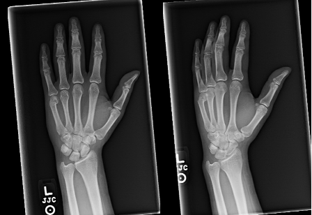
A 27-year-old male presents to the ED with left hand pain after punching a wall. He has pain and swelling on the ulnar side of the dorsal hand. The above hand radiographs were obtained (image courtesy of Mark Hopkins).

This patient has a fracture of the 5th metacarpal neck, otherwise known as a Boxer’s fracture. It is so named because a majority of these fractures come from punching an object [1].

  • Pearl: Unlike most hand and wrist fractures, metacarpal fractures are more likely to occur from axial loading than from a fall onto an outstretched hand (FOOSH) [1].

1. Skin:

  • Closely observe for any breaks on the dorsal surface, especially near the MCP joint, as this can indicate an open fracture which would require operative irrigation, debridement, and antibiotics.

2. Angulation:

  • Observe for any obvious malalignment. Dorsal angulation may cause depression of the MCP joint and disappearance of the normally appearing knuckle [1].

3. Neurovascular:

  • As always, any neurovascular deficits should prompt surgical consultation.
  • Pearl: Given the tight fascial layers of the hand, keep compartment syndrome in mind.

4. Rotational Alignment:

  • Observe by having the patient make a fist and checking for proper finger alignment. Extending lines from the fingers should show eventual convergence at the scaphoid. Any degree of malrotation warrants urgent surgical consultation [3].

Image of hand malrotation

Opinions vary, but most agree that any angulation beyond 30 degrees at the metacarpal neck requires reduction [1].

  • PEARL: Anesthesia can be achieved by an ulnar nerve or hematoma block, with reduction accomplished by applying dorsal pressure with the MCP, PIP, and DIP in flexion.

Patients should be placed in an ulnar gutter splint. They can follow up with a hand specialist as determined by institutional policy as an outpatient in 1 week.

  • Pearl: Studies have shown that patients with less than 60 degrees of angulation can be managed non-operatively and expect a full functional recovery. Cosmetic defects may occur, but are often preferable to surgical intervention if function is kept [4].
  • Pearl: If the fracture is comminuted, significantly angulated, malrotated, or intraarticular, educate the patient the possibility of surgical fixation [4].

References

Looking to bone up in general? Check out the SplintER archives. Want more information on hand radiographs? Check out SplintER Series: Case of a First Metacarpal Fracture or Trick of the Trade: Reducing the metacarpal neck fracture.

  1. Malik S, Herron T, Rosenberg N. Fifth Metacarpal Fractures (Boxer’s Fracture). In: StatPearls [Internet]. Treasure Island (FL): StatPearls Publishing; 2020 Jan. PMID: 29261999
  2. Nakashian MN, Pointer L, Owens BD, Wolf JM. Incidence of metacarpal fractures in the US population. Hand (NY). 2012;7(4):426-430. PMID: 24294164
  3. Yang S, Kim JP. Hand Fractures. J Korean Fract Soc. 2018 Apr;31(2):61-70. DOI: 10.12671/jkfs.2018.31.2.61
  4. van Aaken, J, et al. Fifth metacarpal neck fractures treated with soft wrap/buddy taping compared to reduction and casting: results of a prospective, multicenter, randomized trial. Archives of Orthopaedic and Trauma Surgery: Including Arthroscopy and Sports Medicine. January 2016 136(1):135-142. PMID: 26559192

SAEM Clinical Image Series: Eye Injury

eye

An 11-year-old male presented to a pediatric trauma center following a motor vehicle collision (MVC). He was the restrained front-seat passenger when his vehicle was struck head-on, causing frontal airbag deployment. His primary complaint was pain around his right eye with associated blurry vision. He denied diplopia, pain with extraocular movements, flashers, floaters, or curtains in his vision.

(more…)

SplintER Series: Pain in the Snuff Box

Scaphoid Fracture
 
A 16-year-old male presents to the ED after injuring his wrist during a track meet. The patient was running hurdles when he fell forward, planting his wrist into the ground. The imaging is shown below (courtesy of Dr. Hani Makky ALSALAM, Radiopaedia.org).
Scaphoid fracture (Image 2).

  • Pearl: The scaphoid is the most frequently fractured carpal bone [1,2].
  • Pearl: Fractures occur at the waist, proximal third, and distal portion: 65%, 25%, and 10% respectively [3].

Image 2. Fracture of scaphoid. Case courtesy of Dr. Hani Makky ALSALAM, Radiopaedia.org, rID: 10398 (arrow added by authors).

Occurs when there is an axial load across hyper-dorsiflexed, pronated and ulnar deviated wrists or from a fall on the outstretched hand (FOOSH) [1-3].

Snuff box tenderness, scaphoid tubercle tenderness over the volar aspect of the wrist, and/or positive scaphoid compression test (pain reproduced with an axial load applied through thumb metacarpal) [4-6].

Snuff Box

Image 3. Location of scaphoid tubercle (S) at the base of the thenar eminence (left) and the location of the snuffbox (SB) on the radial aspect of the wrist (right). Images by authors.

Plain film imaging with anterior-posterior, oblique, and lateral views to assess for injury.

  • Pearl: There is also a scaphoid view that is recommended if the department technician is trained. This image is a posterior-anterior view of the scaphoid that is obtained with the wrist in ulnar deviation [7].

Abnormal exam: If not neurovascularly intact or if there is an open fracture, consult orthopedics in the ED.

Identified scaphoid fracture: Thumb spica splint and prompt orthopedic follow-up usually within 1-3 days as though some fractures only require immobilization for treatment; surgery may be required for some fracture patterns [1-3,6].

Suspicion for fracture without radiographic evidence: Place in thumb spica splint and repeat imaging in 14 days to evaluate for occult fracture. If negative again at that time with high clinical suspicion, the patient should have an outpatient MRI [1-3,6].

  • Pearl: Initial imaging can miss 5-20% of fractures [8].

Classic complications include vascular necrosis (AVN), and scaphoid nonunion advanced collapse (SNAC). Associated fractures and dislocation of the surrounding carpal bones, distal radius, ligamentous disruption may be seen as other pathology occurs secondary to a FOOSH [1-4,6].

  • Pearl: AVN is of high concern and directly correlated to the site of fracture. The scaphoid receives blood supply via retrograde flow – the more proximal the fracture, the higher the risk of AVN [1-4,6].
  • Pearl: SNAC occurs when the proximal scaphoid remains attached to the lunate and the distal fragment rotates into flexion. This results in early osteoarthritis between the distal scaphoid and radial styloid, leading to pain and decreased functionality [9].


References & Resources:

For a review of other causes of traumatic wrist pain check out the SplintER archives.

  1. Tada K, Ikeda K, Okamoto S, Hachinota A, Yamamoto D, Tsuchiya H. Scaphoid Fracture–Overview and Conservative Treatment. Hand Surg. 2015;20(2):204-209. PMID 26051761.
  2. Sabbagh MD, Morsy M, Moran SL. Diagnosis and Management of Acute Scaphoid Fractures. Hand Clin. 2019;35(3):259-269. PMID 31178084.
  3. Gupta V, Rijal L, Jawed A. Managing scaphoid fractures. How we do it?. J Clin Orthop Trauma. 2013;4(1):3-10. PMID 26403769.
  4. Basu A, Lomnassey LM, Demos TC, et al: Your Diagnosis? scaphoid fracture. Orthopedics 28:177, 2005. PMID 15751361
  5. Watson HK, Weinzweig J. Physical examination of the wrist. Hand Clin. 1997;13(1):17-34. PMID 9048180.
  6. Stapczynski, JS, Tintinalli, JE. Wrist injuries. In Tintinalli’s emergency medicine: A comprehensive study guide, 8th Edition. New York, NY: McGraw-Hill Education; 2016: 1853-1854
  7. Cheung GC, Lever CJ, Morris AD. X-ray diagnosis of acute scaphoid fractures. J Hand Surg Br. 2006;31(1):104-109.PMID 16257481.
  8. Ashmead D 4th, Watson HK, Damon C, Herber S, Paly W. Scapholunate advanced collapse wrist salvage. J Hand Surg Am. 1994;19(5):741-750. PMID 7806794.
  9. Moritomo H, Tada K, Yoshida T, Masatomi T. The relationship between the site of nonunion of the scaphoid and scaphoid nonunion advanced collapse (SNAC). J Bone Joint Surg Br. 1999;81(5):871-876. PMID: 10530853.
  10.  

Treating Blood Pressure in Intracranial Hemorrhage

hemorrhagic stroke equal podcast

Blood pressure control in the setting of ischemic stroke has a clearly recognized benefit in patient outcomes. The impact of blood pressure control in hemorrhagic stroke is not as well understood. The ACEP E-QUAL Network podcast, a partnership with ALiEM to promote clinical practice improvements, reviewed this topic with Dr. Latha Ganti (University of Central Florida College of Medicine). Dr. Ganti addressed the evidence behind recommended blood pressure targets and the available medications to achieve control. We present highlights from this discussion with Dr. Jason Woods.


What is the goal of blood pressure control in hemorrhagic stroke?

Management of blood pressure in intracranial hemorrhage (ICH) raises questions about the benefit of limiting hematoma expansion while maintaining cerebral perfusion. While it seems intuitive that hypertension should be controlled to limit hematoma expansion, patients with hemorrhagic stroke may be dependent on higher blood pressures for adequate perfusion.

Does lowering blood pressure lead to perihematomal ischemia?

ICH Adapt studies did not show evidence of decreased cerebral blood flow in perihematomal tissue and demonstrated that there is likely preservation of autoregulation which prevents ischemia [1].

Does lowering BP help prevent hematoma expansion and improve outcomes?

The risk of hematoma expansion is highest within the first couple of hours following initial bleeding. Hematoma expansion is clearly associated with worse outcomes. Scoring tools exist to estimate the risk of hematoma expansion. The “spot sign,” seen on source images from a computed tomography angiogram of the brain, suggests an area of dynamic bleeding.

  • ICH ADAPT: no difference in hematoma expansion or clinical outcome with acute blood pressure lowering [2].
  • INTERACT 2: intensive lowering of blood pressure did not result in a significant reduction in mortality or severe disability [3].
  • ATACH 2: intensive lowering of blood pressure did not improve functional outcomes but was associated with increased renal dysfunction [4].

What is the optimal systolic blood pressure (SBP) target?

AHA Guidelines 2015

  • ICH patients with SBP 150-220 mmHg, lower to 14 mmHg is safe
  • ICH patients with SBP > 220 mmHg, aggressive reduction with continuous infusion may be reasonable

So what’s the right thing to do? If data suggests that lowering may not be as beneficial, what should the target blood pressure be?

  • Target SBP 140-160 mmHg is a reasonable target

What medications are preferred for blood pressure control in ICH?

The ideal agent for blood pressure management in ICH would have a quick onset, but short duration, to allow titration.

Recommended first-line:

  • Labetalol
    • Onset < 5 min
    • Duration of effect 2-4 hr
    • IV bolus dose: 20 mg, followed by 20-80 mg every 10 min to a total dose of 300 mg.
    • Infusion dose: 0.5 mg-2 mg/min
    • Avoid in: asthma, COPD, heart failure, AV block
  • Nicardipine
    • Onset 1-2 min
    • Half-life ~ 40 min
    • Infusion dose: 0.5-1 mcg/kg/min, max 3 mcg/kg/min
  • Clevidipine
    • Onset 1-4 min
    • Duration of effect 5-15 min
    • Infusion dose: 1 mg/hr, up to 21 mg/hr, titrate by 2.5 mg/hr every 5-10 min
    • Avoid in: severe aortic stenosis, and lipid metabolism dysfunction or known allergy to eggs or soy (delivered as lipid emulsion)

Available second-line (mostly off-label, not preferred)

  • Esmolol
  • Fenoldopam
  • Hydralazine
  • Enalaprilat

Conclusions

When it comes to blood pressure: keep it simple.

  • Target SBP 140-160 mmHg
  • Top three drugs: Labetalol, Nicardipine, Clevidipine

Although labetalol has common contraindications, it is available as a bolus dose. In a clinical setting where drips may not be readily available, Labetalol can be easier to get.

Interested in more ACEP-EQUAL podcasts?

Listen to the other ACEP E-QUAL podcasts on our Soundcloud account.

References

  1. Butcher K, Jeerakathil T, Emery D, et al. The Intracerebral Haemorrhage Acutely Decreasing Arterial Pressure Trial: ICH ADAPT. Int J Stroke. 2010;5(3):227-233. PMID: 20536619
  2. Butcher KS, Jeerakathil T, Hill M, et al. The Intracerebral Hemorrhage Acutely Decreasing Arterial Pressure Trial. Stroke. 2013;44(3):620-626. PMID: 23391776
  3. Anderson CS, Heeley E, Huang Y, et al. Rapid blood-pressure lowering in patients with acute intracerebral hemorrhage. N Engl J Med. 2013;368(25):2355-2365. PMID: 23713578
  4. Qureshi AI, Palesch YY, Barsan WG, et al. Intensive Blood-Pressure Lowering in Patients with Acute Cerebral Hemorrhage. N Engl J Med. 2016;375(11):1033-1043. PMID: 27276234

SplintER Series: A Case of Inability to Move the Knee

Normal knee radiograph

29-year-old F presents to ED with acute onset knee pain. Reports hearing an audible “pop” after twisting her leg while running down the stairs at home. She explains that her right knee is stuck, and she can neither flex nor extend it. An image is shown below (courtesy of Andrew Murphy, Radiopaedia.org)


(more…)

SAEM Clinical Image Series: Pitching Pain

pitching pain

A twenty-year-old right-handed male presented to the emergency department with a past medical history of right coracoid impingement, and three months of increasing right shoulder pain that became suddenly worse. He had a right shoulder arthroscopy nine months ago and played a full season as his baseball team’s pitcher over the past four months. He endorsed no exacerbating symptoms other than movement and has only taken naproxen over the counter for this pain. He denied any family history of clotting disorders.

(more…)

SAEM Clinical Image Series: Knee Pain

knee

A fifty-six-year-old male with a past medical history of legal blindness and remote right quadricep tendon rupture presents to the emergency department via emergency medical services (EMS) after a mechanical fall, complaining of left knee pain. According to the patient, he is in his regular state of health and was walking with his cane when he had a mechanical fall on the sidewalk after tripping on an unknown object and falling onto his left knee.

The patient did not hit his head, did not lose consciousness, and has no head, neck, or back pain. The patient states that he fell directly onto his knee and felt a popping upon hitting the ground, and remembers all events surrounding the incident. The patient was not ambulatory prior to coming to the emergency department.

(more…)

Go to Top