Tricks of Trade: Benign paroxysmal positional vertigo | Beyond the Basics

Benign paroxysmal positional vertigo

Clinical Case

A 63-year old female presents to your ED with positional dizziness since rising out of bed from a nap this afternoon. She says she had a similar episode in the past and reports, “they took the stones out of my ear by making me lay down and move my head a few times.” Based on your assessment of the patient’s history and physical exam you determine she has peripheral vertigo, likely BPPV. However, despite multiple attempts with the Epley Maneuver, the patient is still symptomatic. What next steps could you consider?

Benign paroxysmal positional vertigo: The basics

Benign paroxysmal positional vertigo (BPPV) is a type of peripheral vertigo caused by a cluster of otoconial fragments that are displaced into the involved semicircular canal. The classic presentation is brief episodes of dizziness reported with position changes, commonly with rolling or arising from bed. The condition is more common in females and with advanced age (>40). BPPV should be differentiated from central vertigo and other types of peripheral vertigo including Meniere’s disease, vestibular schwannoma, vestibular neuritis, and labyrinthitis among others. Displaced otoliths are most commonly located in the posterior or horizontal semicircular canals. The strongest positive predictors of BPPV include dizziness lasting <15 seconds and onset with turning over in bed [1]. Episodes occur more frequently in the ear that is habitually dependent while sleeping [2], most commonly the right ear [3]. Regarding canal involvement, a retrospective review of 253 patients demonstrated the following [4]:

  • 83% Unilateral posterior canal
  • 7% Unilateral horizontal canal
  • 6% Bilateral posterior canals
  • 0% Anterior canal

There exist many different diagnostics and therapeutic positional techniques for addressing BPPV. Below we discuss the commonly taught techniques and several viable alternatives to consider when initial evaluation and/or treatment are unsuccessful.

Posterior Canal


1. Diagnostic: Loaded Dix-Hallpike Test

A Dix-Hallpike test is the most commonly taught and used diagnostic technique. However, providers may consider the “loaded” Dix-Hallpike.

Technique: Flex the patient’s head forward 30° in the same plane as the affected posterior canal for 30 seconds before placing supine with traditional technique. The loaded Dix-Hallpike has increased sensitivity, duration of nystagmus, and severity of symptoms compared to the traditional techniques [5]. Consider using pillow/blankets under the thoracic spine to allow adequate cervical extension as an alternative to hanging the patient’s head over the end of the bed (trick of the trade). Elderly patients with severe kyphosis may need to be tested with the head of the bed tilted downward (Trendelenburg).


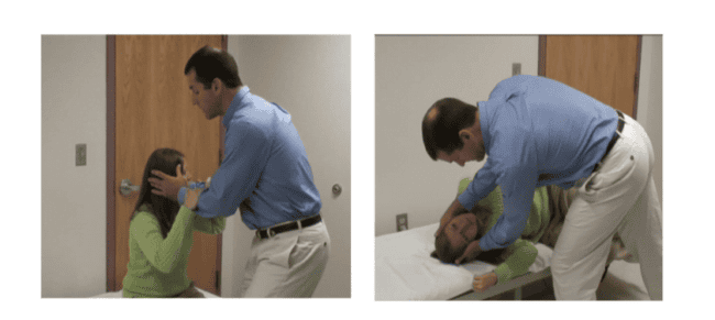
2. Diagnostic: Sidelying Test

This is an alternative to Dix-Hallpike in patients who cannot lie flat, such as with back pain, limited mobility, obesity, or orthopnea. It can be performed on the edge of the bed (often logistically easier in crowded ED rooms than Dix-Hallpike).

Technique: Rotate the head 45° contralateral to the posterior canal being tested. The patient descends to their side which is ipsilateral to the posterior canal being tested. This position is held for 30 seconds. If the patient experiences vertigo and the provider notices nystagmus, the test is positive. A negative test should prompt testing on the other side.


3. Therapeutic: Epley Maneuver

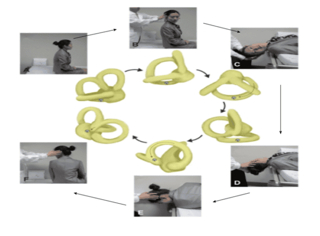
This is the most commonly taught and performed repositioning maneuver. The American Academy of Neurology and American Academy of Otolaryngology has given this technique a Level A Recommendation and clinical benefit demonstrated in a systematic review [6]. Consider using a “chin tuck”, similar to the loaded Dix-Hallpike, for additional success.

Epley Maneuver vertigo

Epley Maneuver


4. Therapeutic: Semont Maneuver

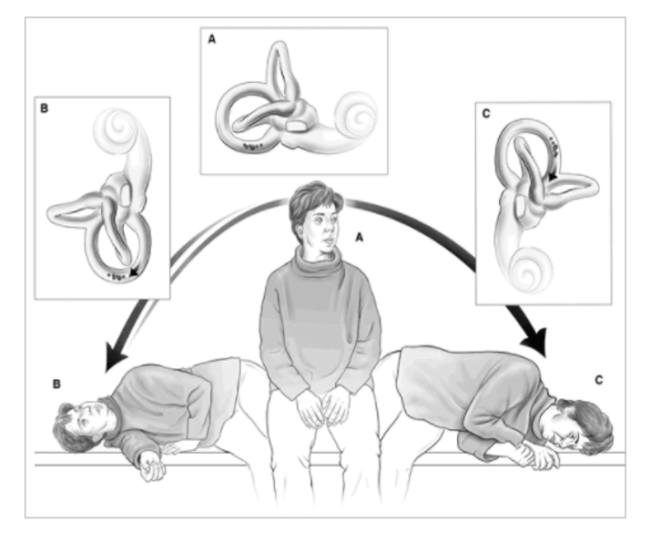
Much like the Epley Maneuver is a continuation of the Dix-Hallpike Test, this therapeutic maneuver is a continuation of the Sidelying Test. The technique for left-sided posterior canalithiasis involves having a seated patient turn their head 45° to the left. The patient then drops their trunk to the right side, with the head turned 45° to the left (facing “up”). This position is held for 30-60 seconds. The patient then quickly sits up and lies down on the left side without stopping in the seated position. The head should still be kept 45° to the left so that the head now faces “down” and into the bed. This position is held for 30-60 seconds. Return the patient to the upright position.


Semont Maneuver vertigo

Semont Maneuver desired otolith movement

Horizontal Canal


1. Diagnostic: Roll Test

The Roll Test should be considered in patients displaying symptoms consistent with BPPV but posterior canal tests (Dix-Hallpike, Sidelying) are negative or appear to demonstrate horizontal nystagmus.

Technique: Have the patient begin by lying supine with the head flexed forward 30°. The provider then rotates the patient’s head rapidly 90° to one side followed by the other side, after re-centering the head. A positive test will involve bursts of nystagmus beating towards the affected ear which are stronger when the affected ear is dependent.


2. Therapeutic: BBQ or Lempert Roll

This repositioning maneuver can be performed as a continuation of the Roll Test and has shown success rates over 90% [7].

Technique: This involves stepwise rotations of the non-tilted head starting in the supine position and ultimately rolling a full 360°, holding each incremental 90° rotation for 30 seconds, starting from the affected to the unaffected side. This  can be repeated 2-4 times until symptoms improve or nystagmus disappears.


3. Therapeutic: Appiani/Gufoni Maneuver

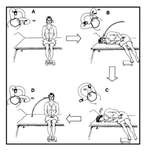
The Appiani/Gufoni Maneuver repositioning maneuver has shown success rates comparable to other techniques in a meta-analysis [8].

Technique: Have the sitting patient descend to their unaffected side, hold this position for one minute or until symptoms subside. Then turning the head 45° towards the bed, holding this position for 1-2 minutes before sitting back up. Repeat until nystagmus is absent.

Appiani/Gufoni Maneuver vertigo

Appiani/Gufoni Maneuver desired otolith movement

Anterior Canal

The same maneuvers can be used to treat both posterior and anterior BPPV (i.e., Epley, Semont). However, there is a paucity of literature given the rarity of this condition. One small study reports success using a “reverse Epley” in 2 of 4 patients [9].

General Guidelines

  1. If your initial therapeutic approach does not work, consider treating the other side as the side of dysfunction can be easily misidentified at first. Serial examinations are often required to confirm BPPV.
  2. Providers should be aware of any underlying spinal or carotid disorders when performing many of the rapid head movements in these patients.
  3. Patients should be observed for a short time immediately after repositioning for signs of possible worsening symptoms and risk of fall [12].
  4. In cases of bilateral BPPV, consider treating the less involved side initially, followed by the more involved side 10-15 minutes later.
  5. Recurrence is common unfortunately despite successful therapeutic intervention. Up to 44% of patients had recurrent symptoms at 2-year follow-up in one study [6].
  6. Patient education: After successful treatment, sleeping slightly elevated or on the uninvolved side may prevent recurrences [10, 11].

Case Resolution

Realizing that you may have mis-identified the side and location of the dysfunction, you perform maneuvers assuming alternative locations for the provoking otoliths. To test for horizontal canal (instead of the more common posterior canal) dysfunction, you perform the roll test and notice nystagmus and worsening symptoms when facing the right side. Consequently, you have the patient perform the Lempert Roll technique, which causes her symptoms to resolve.

While you observe her for 10 minutes, there is no recurrence of her symptoms and she can ambulate without issues. You advise her to sleep on her left side. Outpatient follow-up with a physical therapist, specializing in vestibular disorders, should be strongly considered, especially if the patient is at risk for falls or if responsiveness to treatment was unclear.


The authors would like to extend a special thanks to Jeff Walter PT, DPT, NCS whose in-depth knowledge, experience, and research in the area of vestibular disorders were essential to this post. He is the creator of a FOAM blog: Vestibular Today on vestibular disorders that include many useful resources, diagrams, and videos.

References

  1. Noda K, Ikusaka M, Ohira Y, Takada T, Tsukamoto T. Predictors for benign paroxysmal positional vertigo with positive Dix–Hallpike test. Int J Gen Med. 2011;4: 809. PMID 22162937
  2. Çakir BÖ, Ercan İ, Çakir ZA, Civelek Ş, Sayin İ, Turgut S. What is the true incidence of horizontal semicircular canal benign paroxysmal positional vertigo? Otolaryngology. 2006 Mar; 134(3):451-4. PMID 16500443
  3. Von Brevern M, Seelig T, Neuhauser H, Lempert T. Benign paroxysmal positional vertigo predominantly affects the right labyrinth. J Neurol Neurosurg Psych Res. 2004 Oct 1; 75(10):1487-8. PMID 15377705
  4. Walters J. Geisinger Vestibular & Balance Center. Unpublished data. 2011.
  5. Andera L, Azeredo WJ, Greene JS, Sun H, Walter J. Optimizing Testing for BPPV–The Loaded Dix-Hallpike. J Int Adv Otol. 2020 Aug; 16(2):171. PMID 32784153
  6. Helminski JO, Zee DS, Janssen I, Hain TC. Effectiveness of particle repositioning maneuvers in the treatment of benign paroxysmal positional vertigo: a systematic review. Physical therapy. 2010 May 1; 90(5):663-78. PMID 20338918
  7. Li J, Guo P, Tian S, Li K, Zhang H. Quick repositioning maneuver for horizontal semicircular canal benign paroxysmal vertigo. J Otol. 2015 Sep; 10(3): 115–117. PMID 29937793
  8. Fu W, Han J, Chang N, et al. Immediate efficacy of Gufoni maneuver for horizontal canal benign paroxysmal positional vertigo: a meta-analysis. Auris Nasus Larynx. 2020 Feb 1; 47(1): 48-54. PMID 31151785
  9. Honrubia V, Baloh RW, Harris MR, Jacobson KM. Paroxysmal positional vertigo syndrome. Am J Otol 1999; 20: 465. PMID 10431888
  10. Shigeno K, Ogita H, Funabiki K. Benign paroxysmal positional vertigo and head position during sleep. J Vestib Res. 2012 Jan 1; 22(4):197-203. PMID 23142834
  11. Li S, Tian L, Han Z, Wang J. Impact of postmaneuver sleep position on recurrence of benign paroxysmal positional vertigo. PloS one. 2013 Dec 18; 8(12):e83566. PMID 24367602
  12. Uneri A. Falling sensation in patients who undergo the Epley maneuver: a retrospective study. Ear Nose Throat J. 2005 Feb; 84(2):82-5. PMID 15794543
By |2021-02-17T11:14:27-08:00Feb 17, 2021|Neurology, Tricks of the Trade|

IDEA Series: Handheld Ultrasound for Emergency Medicine Residents Rotating on Cardiology Services

US System

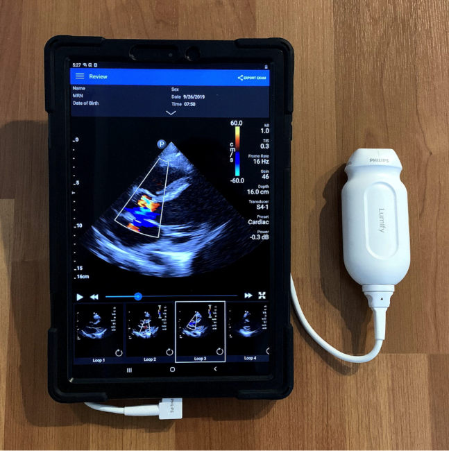
Point-of-care ultrasound (PoCUS) has become an essential skill that emergency medicine (EM) residents learn during their training [1]. Accordingly, most EM programs schedule a block early in residency dedicated to obtaining and interpreting high-quality PoCUS images. Likewise, the ability to efficiently diagnose and manage acute cardiovascular pathologies is a critical aspect of EM, and most EM residents also rotate on a cardiology service to develop these skills. Despite evidence that PoCUS improves the ability of both cardiologists and non-cardiologists to quickly diagnose cardiac disease at the bedside, integration of this relatively novel technology on cardiology services is often limited by lack of PoCUS availability as well as lack of a convenient platform to share recorded images [2]. Equipping EM residents on cardiology rotations with a portable, handheld ultrasound (US) system (Figure 1. Philips Lumify handheld US system with tablet) can enhance the learning of echocardiography acquisition and interpretation while simultaneously providing cardiology teams with clinically actionable information [3]. In addition to improving patient care, performing and interpreting PoCUS from the lens of a cardiologist is a simple yet innovative way to solidify the skills that are crucial to becoming an excellent bedside echocardiographer.

(more…)

By |2021-02-03T21:14:52-08:00Feb 5, 2021|IDEA series, Medical Education, Ultrasound|

SAEM Clinical Image Series: Edema Got Your Tongue?

angioedema

A 57-year-old male presented to the emergency department with a swollen mouth for three hours. He reported never having experienced this before and denied starting any new medications. The patient endorsed a feeling that his mouth was swollen and had difficulty swallowing. The edema had been increasing in size since its onset. He had been drooling for the past hour and endorsed mild pain around the area. He denied any shortness of breath, rash, nausea, vomiting, or other areas of edema. His past medical history included hypertension, diabetes, and allergies, with no known drug allergies. His family history was unknown. His medications included Metformin and Lisinopril.

(more…)

SAEM Clinical Image Series: Sun-burnt Hands and Lips

blistering

A 44-year-old Caucasian male with a past medical history of hepatitis C presents with a complaint of pain, swelling, and skin blistering of his hands. He also notes skin sores on his nose, lower lip, and the tops of his ears. The patient claims that these have become progressively worse since starting work a month ago in outdoor construction. The patient denies the use of medications or illicit drugs and denies any medical allergies. He admits to tobacco use and daily alcohol use. The patient denies any other symptoms.

(more…)

Colchicine Toxicity: A New Threat from COVID-19 Treatments

In the continued fight against COVID-19, a January 22, 2021 press release from the Montreal Heart Institute touted the potential of colchicine, citing results from the COLCORONA trial [1, 2]. We’ve learned to be especially skeptical of any study results reported only via press release before undergoing full peer-review and publication. Nevertheless, the authors claim a non-significant (p=0.08) relative risk reduction of 19% (absolute risk reduction 1.1%) in hospitalizations, mechanical ventilation, and death. Note that the pre-print of the study has still not been peer-reviewed [3]. This study comes on the heels of the much smaller GRECCO-19 study published in June 2020 [4].

Early in 2020, promising results on hydroxychloroquine for treatment of COVID-19 led to a large increase in its use in outpatients and inpatients. It is now known that there is virtually no role for hydroxychloroquine and that this spike in use led to serious toxicity both from therapeutic use and overdose [5, 6]. The same may be anticipated for colchicine. And, if there is a drug that toxicologists fear more than hydroxychloroquine in overdose, it’s colchicine.

5 things to know about colchicine toxicity

1. Colchicine inhibits microtubule formation and function, thereby inhibiting mitosis.

  • It also is a GABA-A antagonist.

2. Acute toxicity occurs in 3 phases.

  • 0-24 hours: Nausea, vomiting, diarrhea, salt and water depletion, and  leukocytosis
  • 1-7 days: Sudden cardiac death (24-48 hours), pancytopenia, acute kidney injury, sepsis, acute respiratory distress syndrome, rhabdomyolysis, and electrolyte imbalance
  • >7 days: Alopecia, myopathy, neuropathy, and myoneuropathy

3. Colchicine levels are not helpful.

  • They also aren’t readily available at most institutions.

4. Hemodialysis doesn’t remove the toxin.

  • Colchicine’s volume of distribution is large.
  • Extracorporeal treatment can be employed if kidney toxicity results from the poisoning.

5. There is no antidote.

  • Management is largely supportive with IV fluids, vasopressors, and colony-stimulating factors.
  • Experimental anti-colchicine Fab fragments are being studied [7].

This 2010 Clinical Toxicology review article provides further information and education on colchicine toxicity.

Want to learn more about EM Pharmacology?

Read other articles in the EM Pharm Pearls Series.

References:

  1. Montreal Heart Institute. Colchicine reduces the risk of COVID-19-related complications. GlobalNewswire website. January 22, 2021. Accessed January 26, 2021.
  2. Montretal Heart Institute. ColCorona. Accessed January 26, 2021.
  3. Tardif J-C, Bouabdallaoui N, L’Allier PL, et al. Efficacy of colchicine in non-hospitalized patients with covid-19. medRxiv. doi: Epub 2021 Jan 27.
  4. Deftereos SG, Giannopoulos G, Vrachatis DA, et al. Effect of colchicine vs standard care on cardiac and inflammatory biomarkers and clinical outcomes in patients hospitalized with coronavirus disease 2019: the grecco-19 randomized clinical trial. JAMA Netw Open.  2020;3(6):e2013136. doi: 10.1001/jamanetworkopen.2020.13136. PMID: 32579195.
  5. Mahan KM, Hayes BD, North CM, et al. Utility of hypertonic saline and diazepam in covid-19–related hydroxychloroquine toxicity. The Journal of Emergency Medicine. doi: 10.1016/j.jemermed.2020.10.048. Epub 2020 Oct 2020. PMID: 33353811.
  6. Chai PR, Ferro EG, Kirshenbaum JM, et al. Intentional hydroxychloroquine overdose treated with high-dose diazepam: an increasing concern in the covid-19 pandemic. J Med Toxicol. 2020;16(3):314-320. doi: 10.1007/s13181-020-00790-8. PMID: 32514696.
  7. Eddleston M, Fabresse N, Thompson A, et al. Anti-colchicine Fab fragments prevent lethal colchicine toxicity in a porcine model: a pharmacokinetic and clinical study. Clinical Toxicology. 2018;56(8):773-781. doi: 10.1080/15563650.2017.1422510. PMID: 29334816.


Go to Top