
High-Dose Nitroglycerin for Sympathetic Crashing Acute Pulmonary Edema

Background
Nitroglycerin (NTG) is an important intervention to consider for patients with Sympathetic Crashing Acute Pulmonary Edema (SCAPE) as it significantly reduces preload, and even modestly reduces afterload with high doses. For acute pulmonary edema in the ED, NTG is often administered as an IV infusion and/or sublingual tablet. Starting the infusion at ≥ 100 mcg/min produces rapid effects in many patients and can be titrated higher as tolerated, with doses reaching 400 mcg/min or greater. Combined with noninvasive positive pressure ventilation (NIPPV) and in some cases IV enalaprilat, patients often turn around quickly, from the precipice of intubation to comfortably lying in bed [1, 2]. But what does the literature say about starting with a high-dose NTG IV bolus followed by an infusion?
Evidence
A 2021 prospective, pilot study of 25 SCAPE patients proposed a clear and systematic protocol (below) for treating these critically ill patients with a combination of high-dose NTG bolus (600 – 1000 mcg over 2 mins) followed by an infusion (100 mcg/min) and NIPPV [3].There were no cases of hypotension after the bolus and 24 of the 25 patients were able to avoid intubation. Additionally, an earlier PharmERToxGuy post summarizes some of the previous studies evaluating the use of a high-dose NTG IV bolus for acute pulmonary edema.
It is important to note that some institutions may not allow IV push NTG or may limit the use of NTG boluses. Providers may then opt to implement dosing strategies such as bolusing from an IV infusion pump or initiating the infusion at a high rate for a short period (e.g., NTG 300 mcg/min for 2-3 minutes) before reducing the rate to a more traditional infusion rate (e.g., 100 mcg/min).
Bottom Line
- A few small ED studies support the use of an initial IV NTG bolus followed by an infusion compared to the infusion alone [1, 2]
- There is a low risk of hypotension following a single IV NTG bolus
- Consider using the following protocol to identify which doses may be best for specific patients based on initial systolic blood pressure

Click for full-sized version [3]
Want to learn more about EM Pharmacology?
Read other articles in the EM Pharm Pearls Series and find previous pearls on the PharmERToxguy site.
References
- Wang K, Samai K. Role of high-dose intravenous nitrates in hypertensive acute heart failure. Am J Emerg Med. 2020;38(1):132-137. doi: 10.1016/j.ajem.2019.06.046. PMID: 31327485.
- Wilson SS, Kwiatkowski GM, Millis SR, Purakal JD, Mahajan AP, Levy PD. Use of nitroglycerin by bolus prevents intensive care unit admission in patients with acute hypertensive heart failure. Am J Emerg Med. 2017;35(1):126-131. doi: 10.1016/j.ajem.2016.10.038. PMID: 27825693.
- Mathew R, Kumar A, Sahu A, Wali S, Aggarwal P. High-dose nitroglycerin bolus for sympathetic crashing acute pulmonary edema: a prospective observational pilot study. The Journal of Emergency Medicine. Published online June 2021:S0736467921004674. doi: 10.1016/j.jemermed.2021.05.011.


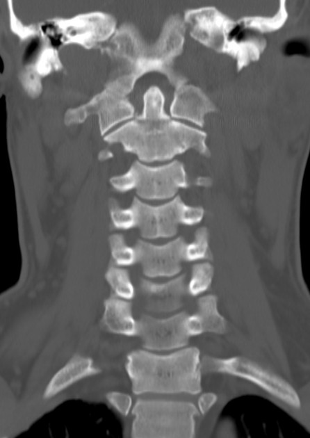
 A 76-year-old female with a history of HTN, TIA, CAD, left CEA, and CKD presented to the emergency department for evaluation of neck bruising and swelling. The patient stated that the night before, she was eating popcorn and choked on a kernel. She states that she coughed to clear her throat and shortly after she developed swelling and bruising to the left side of her neck, which has progressively gotten worse. The patient has a remote history of left carotid endarterectomy and was concerned that her symptoms could be related to the prior surgery. On examination, she had ecchymosis and a hematoma/mass to the left side of her neck without palpable thrill or bruit. A well-healed CEA scar was noted. A CTA of the neck was obtained to determine the source of the ecchymosis/hematoma. What is the diagnosis?