Emergency Medicine factoids on Twitter

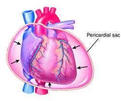
What is a cardiac tamponade? It is a clinical state where pericardial fluid causes hemodynamic compromise. With bedside ultrasonography in most Emergency Departments now, it’s relatively easy to detect a pericardial effusion.
But what we more want to know in the immediate setting is: Is this cardiac tamponade?
You can look for RA systolic or RV diastolic collapse. What if it’s equivocal? How good is the clinical exam and EKG in ruling out a tamponade?
Poor to average, at best. The Beck’s triad of hypotension, distended neck veins, and muffled heart sounds are important to remember … only on tests.
Think about performing a pulsus paradoxus test to see if it’s >12 mmHg. This is a sign of physiologic compromise. Note that the typical cutoff has been 10 mmHg but 12 mmHg is a more specific test.
Adapted from [1]
Go to ALiEM (PV) Cards for more resources.
Thanks to Dr. Hemal Kanzaria for suggesting this JAMA article!
For epistaxis, the classic teaching is to pinch the nose to control the bleeding. A persistent nosebleed often is the result of one’s natural inclination to constantly check if there is still bleeding every few seconds. Applying pressure on-and-off makes it difficult for the bleeding to stop.
To follow-up with Dr. Connolly’s perspective about the Top 10 tips for medical students to rock the EM clerkship rotation, I thought I would post some additional tips. Here are some more pearls:
This means that you should take it upon yourself to make sure that your patient’s care is stellar, addresses key clinical and social issues, and is timely. Constantly check for your patient’s results. Don’t be the last to hear of your patient’s lab or imaging results. Figure out why there are unexpected delays. Address any psychosocial issues which may hamper your patient’s clinical improvement in the ED. (more…)
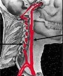
In the setting of blunt trauma, it is easily to overlook a patient’s risk for blunt cerebrovascular injuries (BCVI). These are injuries to the carotid and vertebral arteries. Often they are asymptomatic with the initial injury, but the goal is to detect them before they develop a delayed stroke.
FYI: A simple seat-belt sign along the neck does not warrant a CT angiogram. Patients with higher risk findings such as significant pain, tenderness, swelling, and/or a bruit probably need imaging.
Adapted from [1-3]
Go to ALiEM (PV) Cards for more resources.
It’s that time of year again. When medical students interested in EM are stressing over doing well on their EM rotation.
Here’s a very insightful guest post from Dr. James Connolly, who is a new PGY-1 resident at Hahnemann Hospital in Philadelphia. I’ll write my personal top-10 list next week, from the perspective of a faculty member.
Many MS4 interested in emergency medicine will be starting their EM Sub-I’s in the next few weeks and are naturally wondering what to expect, and how they can be successful, both in terms of getting a strong letter of recommendation, and all while still having a fun and enjoyable rotation. With that in mind, I’d like to present a unique “Top Ten” list, written with the aim of helping the student succeed on his or her upcoming rotation. The list is my own, based on my experiences during three EM rotations last year as a medical student. I’ve also asked a few of my fellow interns for their input on the list, so the list really reflects thoughts of a handful of people who recently successfully matched into emergency medicine.
You are doing a shift in the pediatric ED and you are evaluating a kid with a small bead in her ear. There are a ton different approaches you can use (eg. tissue adhesive glue on a q-tip stick). If the bead is in too deep, blindly trying to adhere the foreign body to the glue is a bit risky. Sometimes applying gentle irrigation might not be enough to wash out the bead. You want to avoid irrigation if you worry about a tympanic membrane rupture.