What’s the Code Dose of tPA?

Suppose you have a patient in whom you highly suspect a pulmonary embolism (PE) that devolves into PEA arrest while awaiting a CT angiogram. Or, what about a patient with an ECG showing clear STEMI that loses pulses?



Suppose you have a patient in whom you highly suspect a pulmonary embolism (PE) that devolves into PEA arrest while awaiting a CT angiogram. Or, what about a patient with an ECG showing clear STEMI that loses pulses?
 There was recently a great study published in the American Journal of Cardiology (2012) by Sharifi et al1, questioning whether we should be considering tPA in patients other than those patients with massive pulmonary embolism (PE)? You know the big “Saddle Embolus” we all fear? Well it turns out this is only about 5% of all PEs.
Should we be considering tPA in patients with sub-massive PEs?

For many of us in academia and medical education, we accomplish a tremendous amount of work outside of the workplace. This can be in our home office, on the public transit system, or in the library.
Interestingly, creative cognition occurs best with a moderate amount of ambient noise (not too much and not too little), according to a 2012 article from Journal of Consumer Research.
(more…)
It’s all about luck, opportunity, and timing.
I will be releasing the blog’s first ever Paucis Verbis (PV) native app this year. After a few years of brainstorming and lots of reader inquiries about an app, we were approached by two different app-building companies in the same week. I’m incredibly humbled to be approached by organizations, who can see the potential of these pocket cards (which started as actual index cards while I was in residency). It is an incredibly exciting time to be in the world of education and social media!
I need your help with step 1.
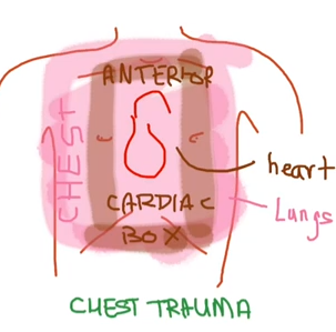
In the third and fourth video in the Trauma series (first two videos = primary and secondary survey), Dr. Rahul Patwari discusses about chest trauma, divided into blunt and penetrating mechanisms. What is “the box” that everyone keeps referring to?
 Pulmonary embolism (PE) can be a deadly disease and one of the most challenging diagnosis to make in a pregnant patient. Patients may present with signs and symptoms that might also be present in a normal uncomplicated pregnancy. Even in nonpregnant patients, the diagnosis of venous thromboembolism (VTE) such as PE can be quite challenging.

Paitents can be a challenge when trying to obtain peripheral IV access. The vein may be collapsed from dehydration or scarred because of IV drug use or repeated cannulation. Before thinking about an ultrasound-guided deep vein IV or a central line, take a look at the external jugular (EJ) vein.