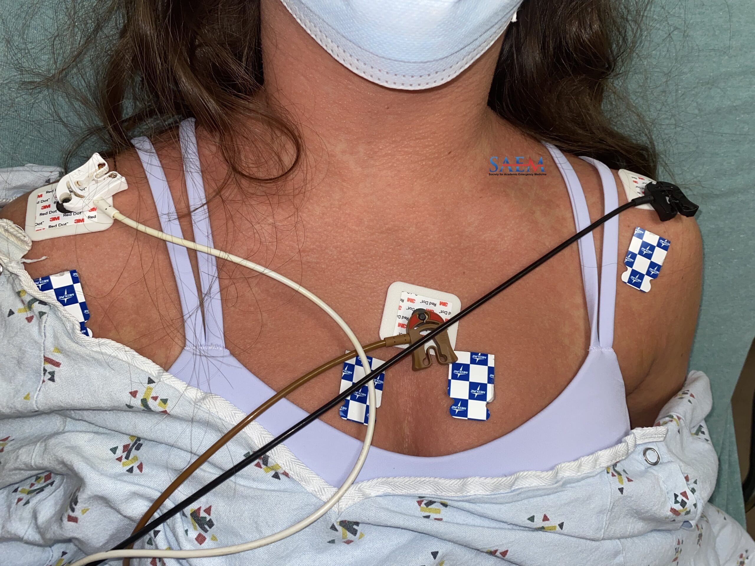
SAEM Clinical Images Series: Face and Chest Rash

chicken pox

A 23-year-old female with a past medical history of asthma presented with a rash that began five days ago on her face and spread to her chest. The lesions are painful and pruritic, spreading slightly to her extremities. She noted a slight sore throat and nasal congestion. She denied any known fever and had no known vaginal or oral lesions. She has a 5-year-old daughter at home with no known symptoms. She is sexually active with one male partner who has no rash or illness. She is vaccinated for COVID-19. She is unsure of childhood illnesses and believes she was never properly immunized as a child in Central America.

Vitals: BP 110/55; Temperature 37°C; pulse ox 97%

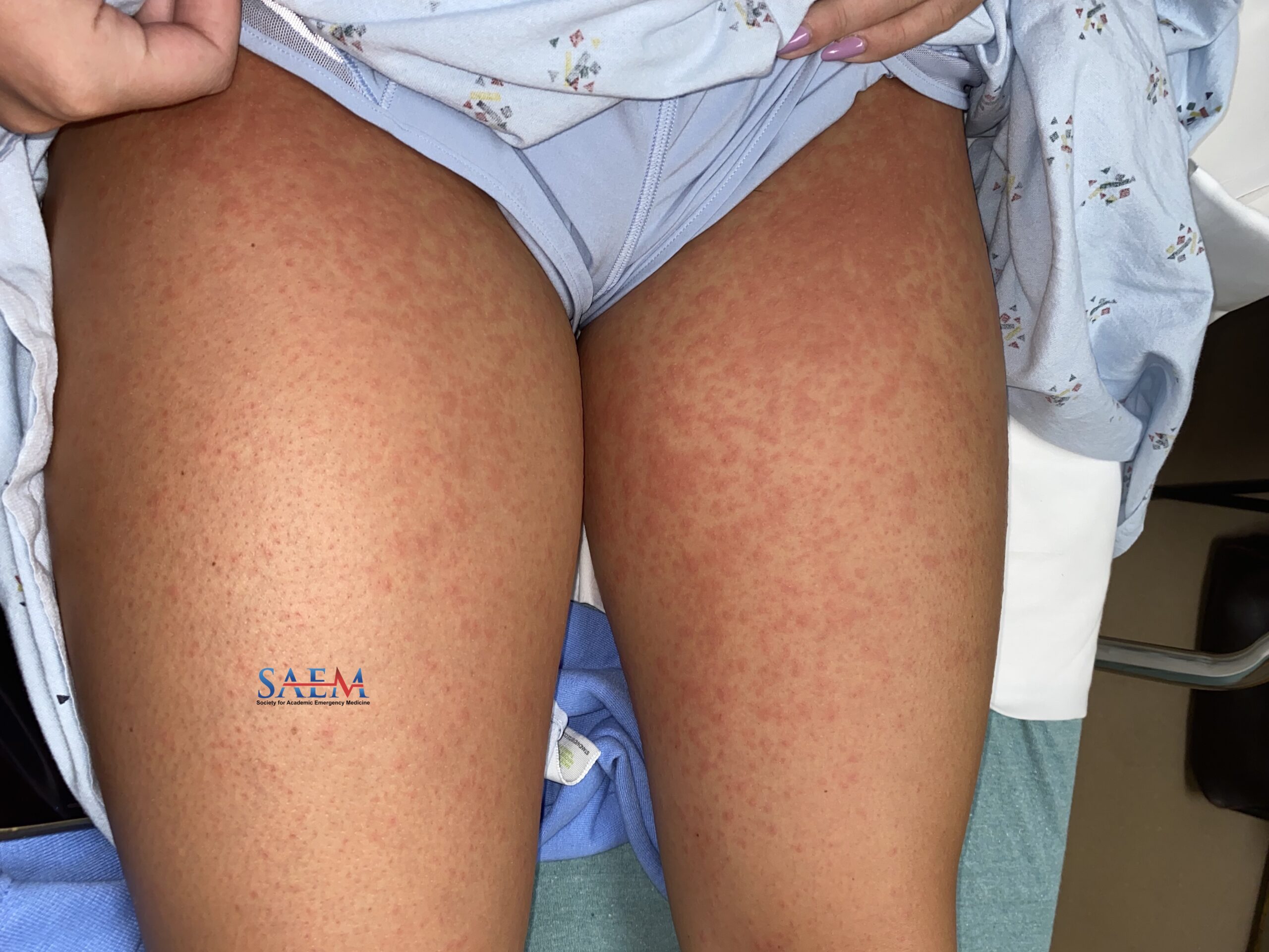
Skin: Face, thorax, and extremity papules are noted with an erythematous base, some vesicular, others with occasional crust or scabs. No dermatomal distribution. There is relative sparing of extremities with more lesions noted on the trunk and face. Negative Nikolsky sign. Otherwise, no other findings on physical examination.

Non-contributory

Herpes varicella-zoster, or chickenpox, is a viral infection transmitted by airborne droplets and direct contact. Before immunization, 90% of cases occurred in children. While primary infection in children is generally benign, adult and infant infections can have severe complications including encephalitis and pneumonia. Erythematous vesicular lesions appear in successive crops typically starting on the face and spreading to the trunk. Extremities usually have more minor involvement with sparing of palms and soles. Vesicles progress quickly to crusted erosions in 8-to-12-hour periods. Treatment should focus on symptomatic care.

Antivirals can slow the severity of the course if given within 24 hours of the onset. Severely immunocompromised patients should receive acyclovir 10 mg/kg IV every 8 hours for 7 to 10 days.

Take-Home Points

  • Chickenpox presents with several “crops” of lesions: papules, vesicles, and scabs.
  • Adult primary disease is not common and is more severe than pediatric illness.
  • Antivirals are not indicated after the first day of illness in immunocompetent adults with mild to moderate disease.

  • Gomez-Gutierrez AK, Flores-Camargo AA, Casillas Fikentscher A, et al. Primary varicella or herpes zoster? An educational case report from the primary care clinic. Cureus Apr 2022;14(4):e23732.
  • Hughes CM, Liu L, Davidson WB, et al. A Tale of Two Viruses: Coinfections of Monkeypox and Varicella Zoster Virus in the Democratic Republic of Congo. Am J Trop Med Hyg. 2020 Dec 7;104(2):604-611. doi: 10.4269/ajtmh.20-0589. PMID: 33289470
  • Kennedy PGE, Gershon AA. Clinical Features of Varicella-Zoster Virus Infection. Viruses. 2018 Nov 2;10(11):609. doi: 10.3390/v10110609. PMID: 30400213
  • Wolff K, Johnson RA Saavedra AP. Fitzpatrick’s Color Atlas of Clinical Dermatology. 7th ed. McGraw Hill. 2013:673-675.

SAEM Clinical Images Series: Utility of Bedside Ultrasonography

fitz-hugh-curtis

A 24-year-old G1P0010 female with a PMHx of ovarian cyst (unknown laterality) and emergency contraceptive use 3 months prior presented with sudden onset abdominal pain (upper > lower) that awoke her from sleep four hours prior to presentation with associated nausea and mild lower back pain. The pain is 10/10, sharp, stabbing, and diffuse. Additionally, she reported trace white vaginal discharge at baseline. No acute increase. She had intermittent vaginal bleeding since contraception use over the past two months, which has now resolved. She denied fever, chills, vomiting, chest pain, shortness of breath, diarrhea, or constipation. No pertinent surgical history.

Constitutional: Uncomfortable. Appearing to be in acute pain.

Cardiovascular: Tachycardia. Regular rhythm and normal heart sounds.

Pulmonary: No respiratory distress. Breath sounds normal.

Abdominal: Diffusely tender abdomen with voluntary guarding, otherwise soft. Normoactive bowel sounds. Negative Murphy’s sign.

Pelvic: Scant white vaginal discharge and CMT. No vaginal bleeding, lacerations, or external lesions.

Neurologic: A&O x 3

WBC: 18.9 k/uL

Hgb: 10.5 g/dL

BMP, lipase, Alk phos/Bili/ALT/AST, PT/PTT, and lactate: Unremarkable

Serum HCG: Negative

Urinalysis (UA): Unremarkable

COVID: Negative

An ideal RUQ ultrasound visualizes the liver, Morrison’s pouch, superior and inferior poles of the right kidney, and diaphragm in the coronal plane. Here, we see a thickened hepatic capsule, septations, and trace ascites.

Fitz-Hugh-Curtis syndrome (FHCS) is characterized by perihepatitis in the setting of pelvic inflammatory disease (PID). It traditionally presents with right upper abdominal pain with associated nausea, vomiting, and fever in women of childbearing age. While overall considered a rare manifestation of PID, the true incidence of FHCS is poorly defined in the literature [1]. The pathophysiology of spread is also poorly understood. It is speculated that bacteria (N. gonorrhoeae, C. trachomatis) travel to the liver via blood, lymphatics or peritoneal fluid, causing perihepatitis [1]. Diagnosing FHCS poses a diagnostic challenge to clinicians. Traditionally, the diagnosis is made via laparoscopic exploration of the abdomen with visualization of the characteristic “violin-string” adhesions, with growing evidence also supporting the use of contrast-enhanced CT [1]. Limited evidence exists to support the use of ultrasonography in diagnosing FHCS. One case report published in 1993 used RUQ abdominal ultrasound to identify septations (violin-string adhesions) and ascites to ultimately diagnose FHCS, later confirmed by serologic and operative evidence [2]. Another case report from 2018 used ultrasonography to identify a thickened hepatic capsule in an 18-year-old female with RUQ pain, later confirming FHCS by CT without the need for laparotomy [3]. While more research is needed, identification of FHCS via bedside ultrasonography in the emergency setting followed by appropriate antibiotic therapy can be an effective approach to FHCS, ideally reserving laparoscopy only for lysis of adhesions in refractory cases.

Take-Home Points

  • RUQ abdominal ultrasound findings of a thickened hepatic capsule, ascites, and septations should raise suspicion for Fitz-Hugh-Curtis syndrome in the emergency setting.

  • Moon, Y.H., Kim, J.H., Jeong W.J., Park, S.Y. Ultrasonographic findings in Fitz-Hugh-Curtis syndrome: a thickened or three-layer hepatic capsule. Yeungnam Univ J Med 35(1), 127-129 (2018).
  • Theofanakis, C.P., Kyriakidis, A.V. Fitz-Hugh–Curtis syndrome. Gynecol Surg 8, 129–134 (2011). https://doi.org/10.1007/s10397-010-0642-8
  • van Dongen PW. Diagnosis of Fitz-Hugh-Curtis syndrome by ultrasound. Eur J Obstet Gynecol Reprod Biol. 1993 Jul;50(2):159-62. doi: 10.1016/0028-2243(93)90181-b. PMID: 8405645.

SAEM Clinical Images Series: Pediatric Genitourinary Bleeding

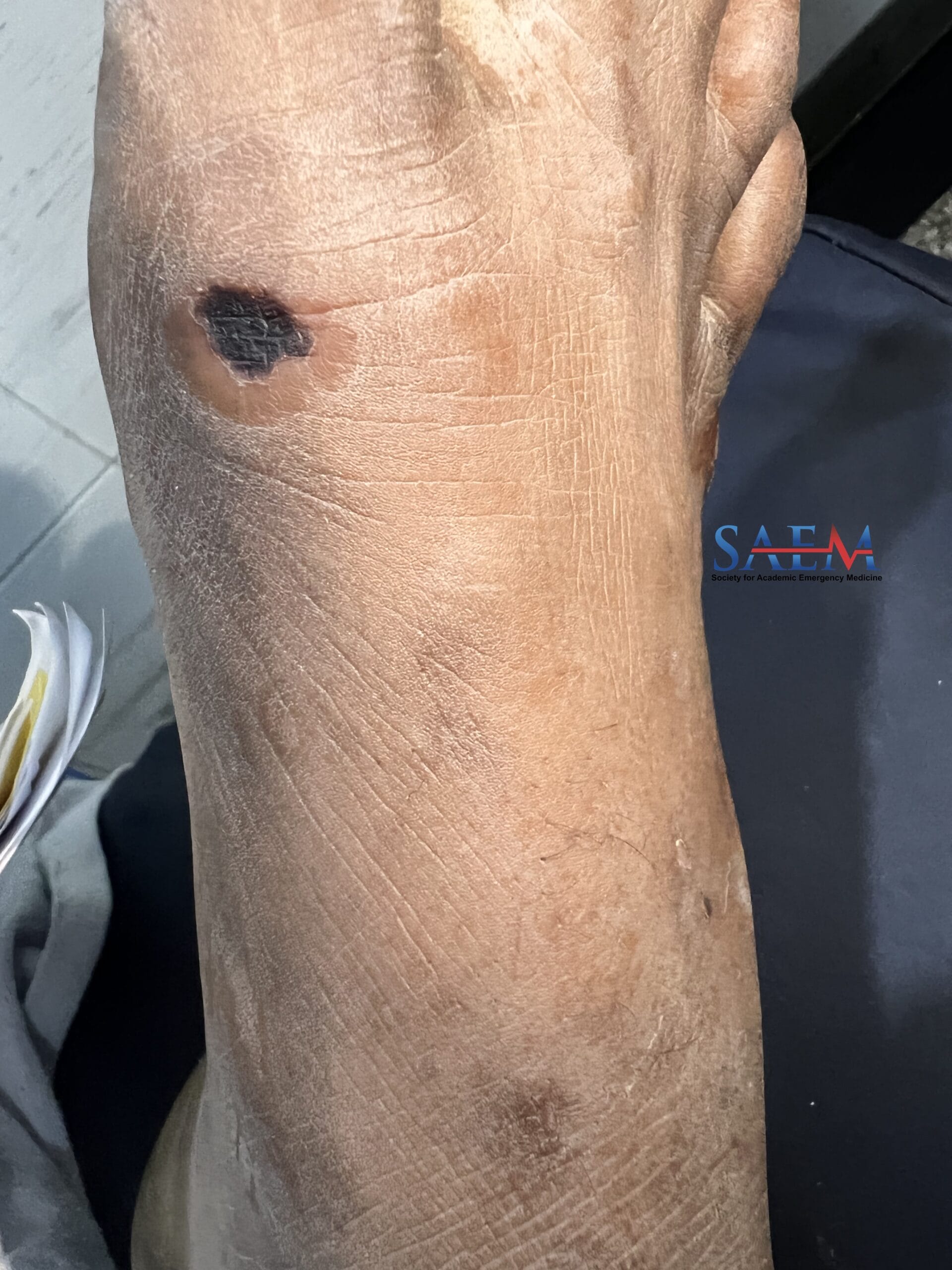
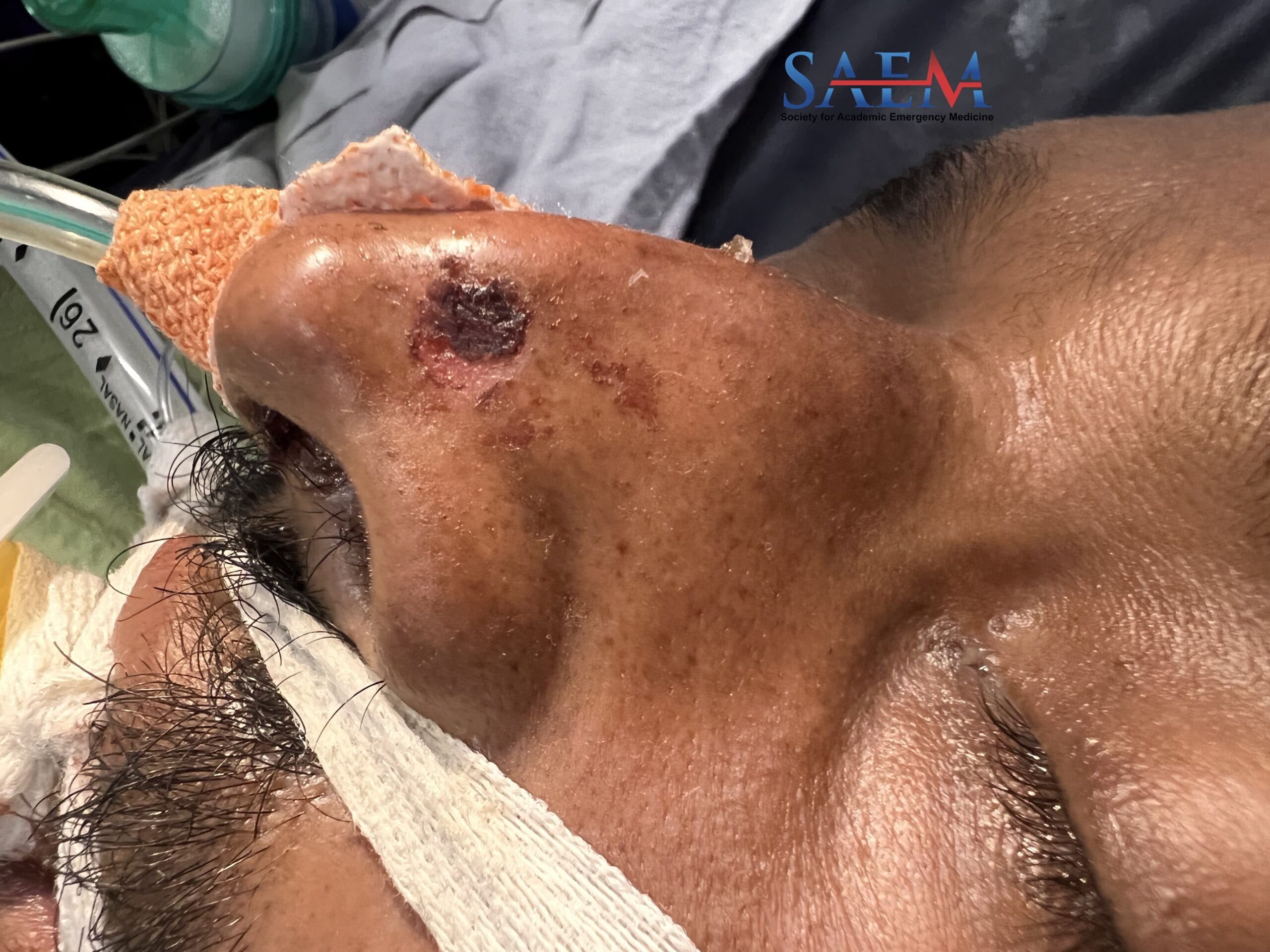
A 4-year-old female with no significant past medical history is brought to the Emergency Department by her grandmother for concern for two days of progressive vaginal bleeding. The grandmother first noted blood in the patient’s underwear the previous morning when she was helping the patient wipe and she noticed it again prior to arrival, this time saturating the patient’s underwear. There is no history of any recent falls or trauma, abdominal pain, pain to the vagina, dysuria, prior incidents of vaginal bleeding, or any noticeable behavioral changes per the grandmother. The patient lives at home with her mother but has been at her grandmother’s house for the past four days (the household consists of female cousins, grandmother, and grandfather). Of note, the patient’s father took her to a trampoline park with her younger sister two days ago.

Vitals: BP 95/68; HR 96; RR 24; 98% on room air; Temp 36.2°C; Wt 18.2 kg

General: Well-appearing 4-year-old female acting appropriately with grandmother and mother at the bedside.

Abdomen: Soft, nontender, nondistended.

Genitourinary: Normal appearing external genitalia without any skin tears/lacerations. Vaginal exam: Slow oozing bleed noted with round “doughnut” shaped tissue protruding at the vaginal opening.

Hemoglobin: 12.2 g/dL

Urinalysis (clean catch): Blood: Large; >200 RBCs, Ketones: 20, Nitrite: Negative, Leukocytes: Moderate, 19 WBCs

FSH/LH/Testosterone: Within normal limits

Urethral prolapse is a rare condition occurring in prepubertal female pediatric patients. It often presents to the emergency department with complaints of vaginal bleeding, difficulty urinating, or dysuria. The most common predisposing factors to this condition include obesity, cough, trauma, constipation, or a history of any activity that causes a sudden recurrent increase in pelvic pressure, such as a trip to the trampoline park as was the case in this patient [1]. On physical examination, urethral prolapse appears as an annular-like mucosal mass with a central dimple located between the labia majora on examination [1]. Initial treatment is medical management with topical estrogen cream in conjunction with Sitz baths and outpatient follow-up with pediatric urology or gynecology. However, persistence of the prolapse or necrosis of the distal urethra often warrants emergent pediatric urology consultation [2,3]. Recurrent cases or cases refractory to medical management will often require surgery. The patient in this case was treated with a 4-week estrogen cream taper. The patient followed up with pediatric gynecology without further complication or need for further intervention.

Child Abuse, Vaginal Trauma, Malignancies (ie: sarcoma botryoides), Infection, Vaginal foreign body, Urethral Prolapse, Precocious puberty, Hypothyroidism, and Exogenous hormone

Take-Home Points

  • Consider urethral prolapse in any prepubertal female who presents to the Emergency Department with a triage complaint of vaginal bleeding.
  • Treatment for urethral prolapse is typically conservative with topical estrogen cream and prompt follow up with pediatric urology or gynecology.
  • Always keep a broad differential for prepubertal pediatric patients with genitourinary and vaginal bleeding complaints
  • HHillyer S, Mooppan U, Kim H, Gulmi F. Diagnosis and treatment of urethral prolapse in children: experience with 34 cases. Urology. 2009 May;73(5):1008-11. doi: 10.1016/j.urology.2008.10.063. Epub 2009 Mar 13. PMID: 19285715.
  • Laufer M, Emans S. Overview of vulvovaginal conditions in the prepubertal child. Uptodate.com. https://www.uptodate.com/contents/overview-of-vulvovaginal-conditions-in-the-prepubertal-child. Published 2021. Accessed January 5, 2022.
  • Teach S. Evaluation of vulvovaginal bleeding in children and adolescents. Uptodate.com. https://www.uptodate.com/contents/evaluation-of-vulvovaginal-bleeding-in-children-and-adolescents#H11716993. Published 2021. Accessed January 5, 2022.

By |2023-11-12T14:19:13-08:00Nov 17, 2023|Ob/Gyn, Pediatrics, SAEM Clinical Images|

SAEM Clinical Images Series: More Than Skin Deep

skin

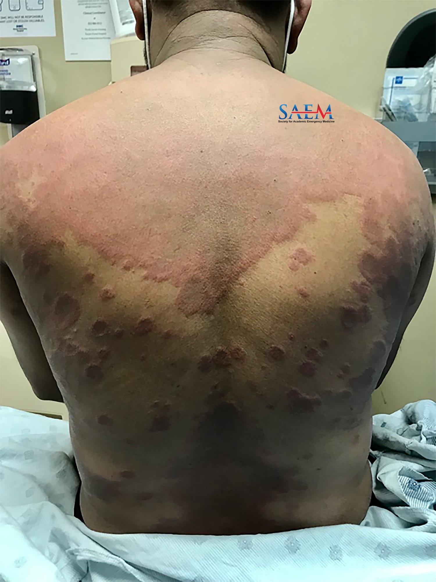
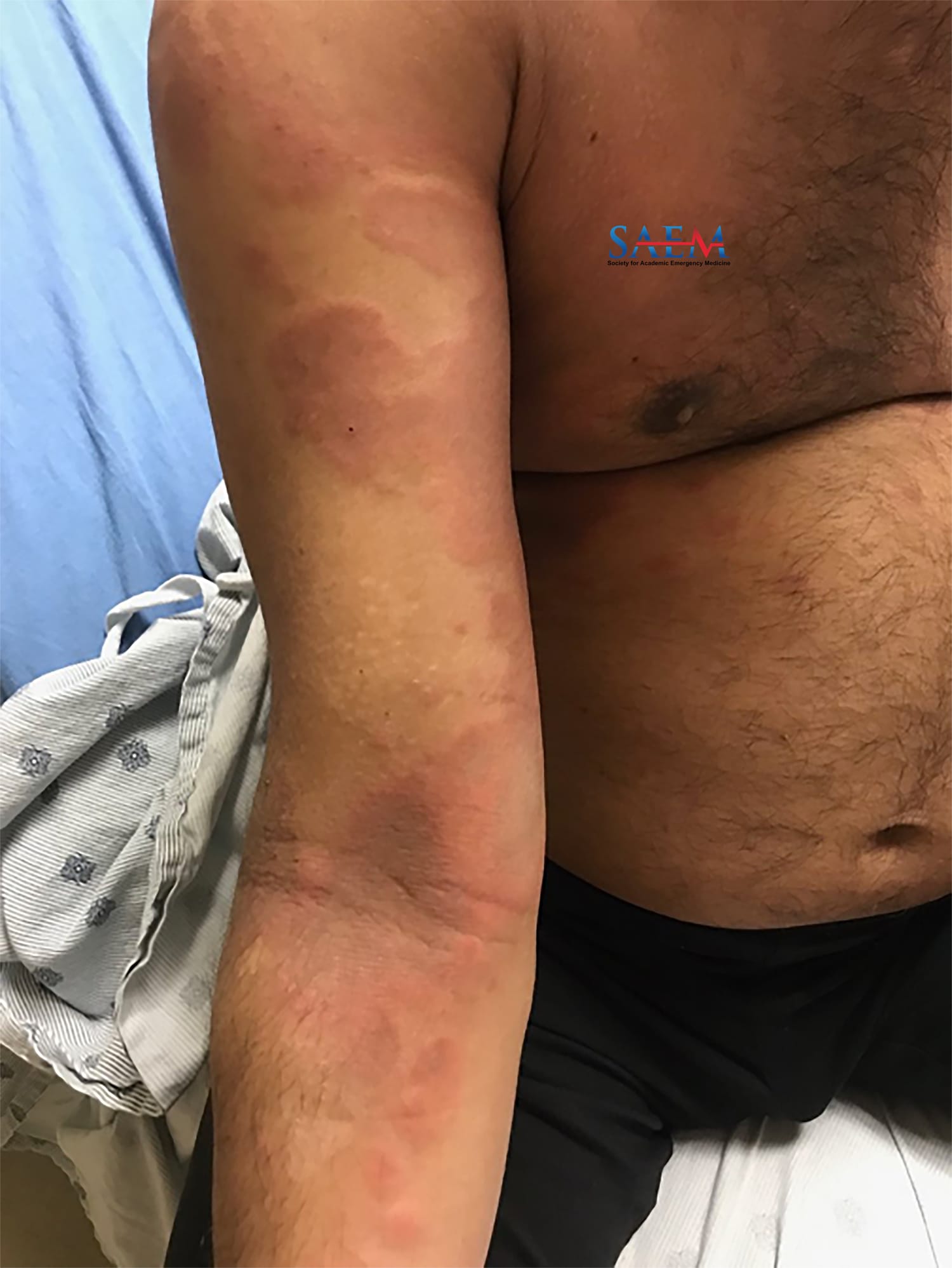
A 57-year-old female college counselor living in the northeastern United States with no PMH presented for evaluation of rash, joint pain, and dyspnea for the past three weeks. The patient first noticed the rash on her upper back, describing it as being itchy. The rash then spread to her face, scalp, and thighs. Two weeks ago, she noticed swelling in her hands and had a gradual onset of dyspnea on exertion. The patient has pain in her hands and when moving her fingers. She denied fever, cough, chills, chest pain, headache, vision changes, focal weakness, abdominal pain, nausea, vomiting, and diarrhea. She denied recent travel, sick contacts, significant time spent outdoors, known tick bites, new medications, and changes in her diet. She has never had a rash like this before.

Vitals: BP 110/70; HR 86 BPM; RR 18 breaths/min; T 37°C; SpO2 96% on RA

Skin: Warm and dry. There is a macular, violaceous rash to the upper back and upper thighs. The patient’s hands are slightly edematous with nontender papules on the palmar aspect of the hands.

CV: Heart sounds are normal. No jugular venous distention or lower extremity edema.

Lungs: There are faint bibasilar rales heard on auscultation of the chest.

Extremities: Full ROM of the joints and there are no bony deformities. The patient does not have muscular tenderness.

Neuro: Within normal limits; muscle strength 5/5 in all four extremities.

CBC w/ differential: Normal

BMP: Na 142, K 3.8, Cl 106, HCO3 24, BUN 16, Cr 0.44, Glu 112, Ca 8.5, Mg 2.2, Phos 3.4

LFT’s: AST 105, ALT 76, ALP 68, Tbili 0.2 Alb 3.7

PT/PTT/INR: 12.0s/32.2/1.04

D-dimer: 347

ESR: 62

CRP: 0.75

Ferritin: 625

CK: 195

LDH: 276

Procalcitonin: undetectable

Amyopathic dermatomyositis – specifically anti-melanoma differentiation-associated gene 5 (anti-MDA5) positive dermatomyositis as determined by subsequent inpatient auto-immunological workup. Compared to other dermatomyositides, anti-MDA5 positive dermatomyositis is characterized by an absence of traditional muscular involvement. Additionally, patients can present with respiratory symptoms related to interstitial lung disease (ILD). One phenotype of this condition is associated with a rapidly progressive ILD, but respiratory involvement may be delayed years after the initial symptoms are noticed. The patient’s clinical images demonstrate a macular, violaceous rash in the “shawl sign” and “holster sign” distribution patterns typical of dermatomyositides. Palmar papules (not to be confused with Gottron’s papules which are found on the dorsal surface of the metacarpophalangeal and interphalangeal joints) are fairly specific for anti-MDA5 positive dermatomyositis

There are no specific guidelines for treating anti-MDA5 positive dermatomyositis. Patients are typically started on a high-dose steroid regimen. A rheumatology consult should be obtained to determine if the patient would benefit from treatment with immunosuppressants. Given her complaints of dyspnea, the patient should undergo a non-contrast CT of the chest to evaluate for evidence of scarring or pulmonary fibrosis.

Take-Home Points

  • Anti-MDA5 positive dermatomyositis is associated with rapidly progressive ILD has a poor prognosis.
  • This rare form of dermatomyositis should be suspected if the patient has respiratory complaints in addition to the hallmark cutaneous findings commonly observed in all types of dermatomyositides. Palmar papules are fairly specific for anti-MDA5 positive dermatomyositis. It often lacks typical historical and physical features of muscular weakness.
  • Treatment involves high-dose corticosteroids and consideration of immunomodulator therapy.

  • Allenbach Y, Uzunhan Y, Toquet S, Leroux G, Gallay L, Marquet A, Meyer A, Guillaud C, Limal N, Gagnadoux F, Hervier B, Borie R, Deligny C, Terrier B, Berezne A, Audia S, Champtiaux N, Devilliers H, Voermans N, Diot E, Servettaz A, Marhadour T, Castelain V, Humbert S, Blanchard-Delaunay C, Tieulie N, Charles P, Gerin M, Mekinian A, Priou P, Meurice JC, Tazi A, Cottin V, Miyara M, Grange B, Israël-Biet D, Phin-Huynh S, Bron C, De Saint Martin L, Fabien N, Mariampillai K, Nunes H, Benveniste O; French Myositis Network. Different phenotypes in dermatomyositis associated with anti-MDA5 antibody: Study of 121 cases. Neurology. 2020 Jul 7;95(1):e70-e78. doi: 10.1212/WNL.0000000000009727. Epub 2020 Jun 2. PMID: 32487712; PMCID: PMC7371381.
  • Nombel A, Fabien N, Coutant F. Dermatomyositis With Anti-MDA5 Antibodies: Bioclinical Features, Pathogenesis and Emerging Therapies. Front Immunol. 2021 Oct 20;12:773352. doi: 10.3389/fimmu.2021.773352. PMID: 34745149; PMCID: PMC8564476.

Go to Top