


Significance of a traumatic pneumothorax or hemothorax found only on CT imaging
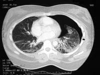 A 32 year old woman arrives in your emergency department after being in a motor vehicle collision where she was the seat-belted driver. She undergoes chest CT imaging despite a negative chest x-ray because of her ongoing anterior chest wall diffuse tenderness. You discover a small 10% pneumothorax (PTX), but no other associated thoracic injuries. Should you place a tube thoracostomy (chest tube)? Should this patient be admitted to the hospital? A 2019 Annals of Emergency Medicine paper by the NEXUS Chest research group tackles these questions.1
 Emergency Medicine (EM) residents are expected to be familiar with and competent in performing a wide number of procedures, including rare ones such as performing an escharotomy in a patient with severe burns. Unfortunately, there is a paucity of readily available simulation models to facilitate practice of this rare yet potentially life-saving skill.
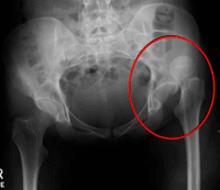 Welcome to Leg Day #2 of the
 A patient presents to your ED with an all too common complaint – chest pain. After a focused history and physical exam, you have an extremely low clinical suspicion for thoracic aortic dissection, pulmonary embolism, pneumonia, pneumothorax, pericarditis/myocarditis, and Boerhaave’s syndrome. When the labs (including a troponin), an ECG, and chest x-ray yield normal results, questions often arise. Can you discharge her with a single troponin if she is low risk? How do you define low risk? And lastly, does she need urgent provocative testing after discharge?
 Accidental hypothermia is a life threatening condition that can lead to a challenging resuscitation. The very young, old, and intoxicated patient are at high risk to developing hypothermia, even in temperate climates. The pathophysiologic changes from hypothermia make the standard ACLS approach insufficient to care for the hypothermic patient. This article will discuss the physiology of hypothermia and how you should alter your approach in the hypothermic patient, including early consideration of extracorporeal membrane oxygenation (ECMO).