Paucis Verbis card: Cervical spine imaging rules
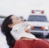 There is constant debate on whether to image the cervical spine of blunt trauma patients. Fortunately, there are two clinical decision tools available to help you with your evidence-based practice.
The NEXUS and Canadian C-spine Rules (CCR) are both validated studies which both quote a high sensitivity (over 99%) in detecting clinically significant cervical spine fractures. Both studies primarily used plain films in evaluating their patients.
| Sensitivity (%) | Specificity (%) | |
| NEXUS | 99.6 | 12.6 |
|---|---|---|
| CCR | 99.4 | 45.1 |
NEXUS
National Emergency X-radiography Utilization Study
A patient’s neck can be clinically cleared safely without radiographic imaging if all five low-risk conditions are met:
- No posterior midline neck pain or tenderness
- No focal neurological deficit
- Normal level of alertness
- No evidence of intoxication
- No clinically apparent, painful distracting injury*
* Defined as “a condition thought by the clinician to be producing pain sufficient to distract the patients from a second (neck) injury. Examples may include, but are not limited to the following:
- Long bone fracture,
- A visceral injury requiring surgical consultation,
- A large laceration, degloving injury, or crush injury,
- Large burns, or
- Any other injury producing acute functional impairment
Physicians may also classify any injury as distracting if it is thought to have the potential to impair the patient’s ability to appreciate other injuries.”
Canadian C-spine Rules (CCR)
The basic approach in this flow-chart is to (1) make sure that the patient meets the same inclusion criteria as in the CCR study. Then (2) determine if there are high-risk findings. If so, go directly to imaging. (3) If there are no high-risk findings, check to see if the patient qualifies as a low-risk candidate where you might be able to clinically clear the c-spine without imaging. (4) If the patient is neither high or low risk, then the patient is moderate risk and requires imaging. Here’s a flow chart that I made to help you remember:
Go to ALiEM (PV) Cards for more resources.
Note: Many emergency physicians go straight to CT imaging for patients with neck tenderness and moderate/high risk findings. I personally rarely use the CCR algorithm because I can rarely remember all of the criteria. NEXUS is nice because of its simplicity. Where the CCR algorithm IS helpful is in clinical clearance of the low-risk patient with neck pain. I’ve cleared many patients who self-present with a whiplash mechanism (simple rear-end motor vehicle crash) and diffuse neck pain. By NEXUS criteria, you’d have to image them because they have neck tenderness. By CCR criteria, if they can actively rotate their neck 45 degrees left and right, they don’t have a clinically significant c-spine injury. No imaging needed.

 Dyphagia is a disorder of swallowing. It actually occurs in up to 10% of adults older than 50 years old. How can you determine the most likely causes for dysphagia? The secret is to obtain a thorough history and using the algorithm below, which I find really helpful from a review article in American Family Physician.