SAEM Clinical Images Series: When it is Not Just a Knot

knot

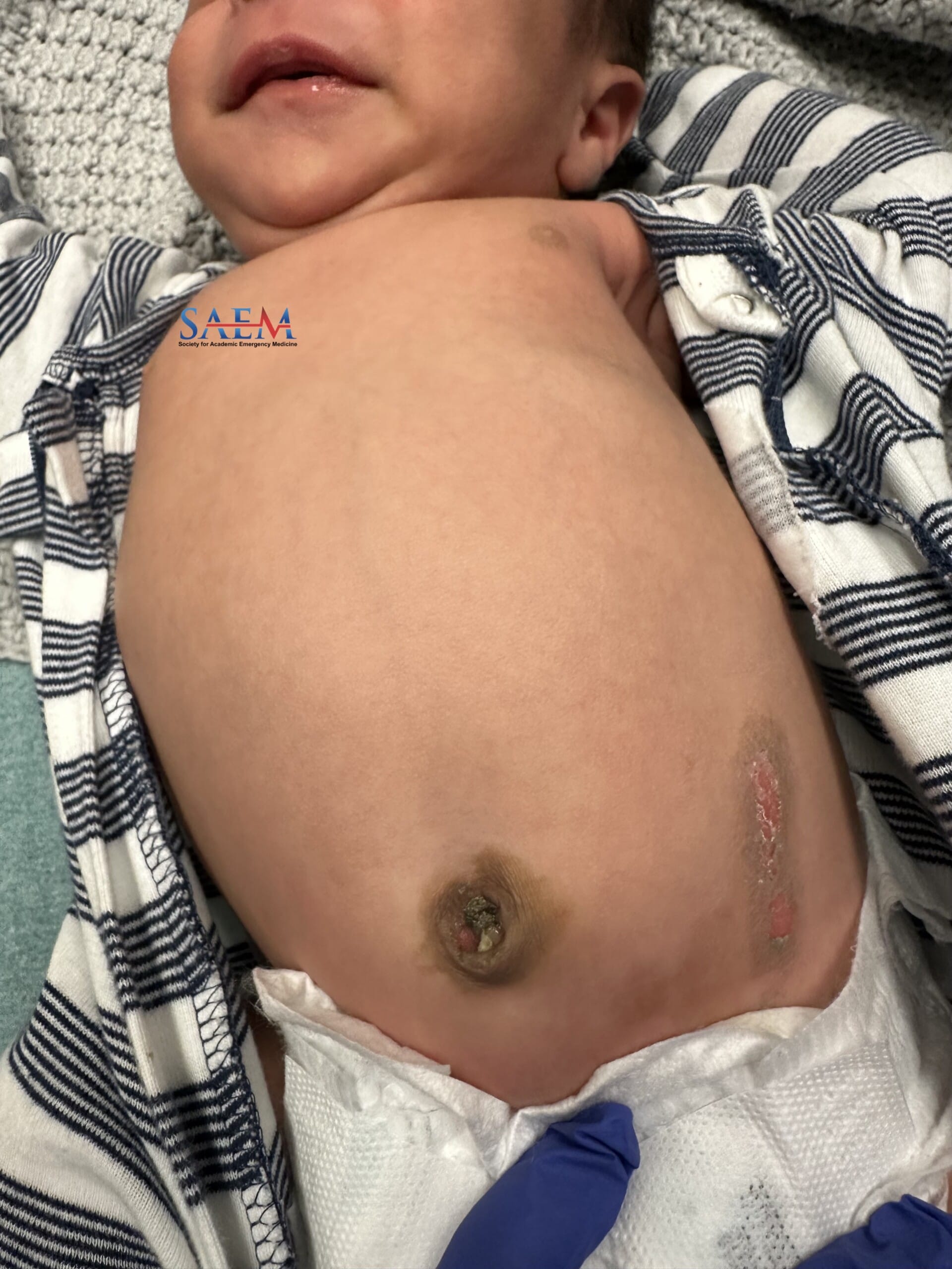
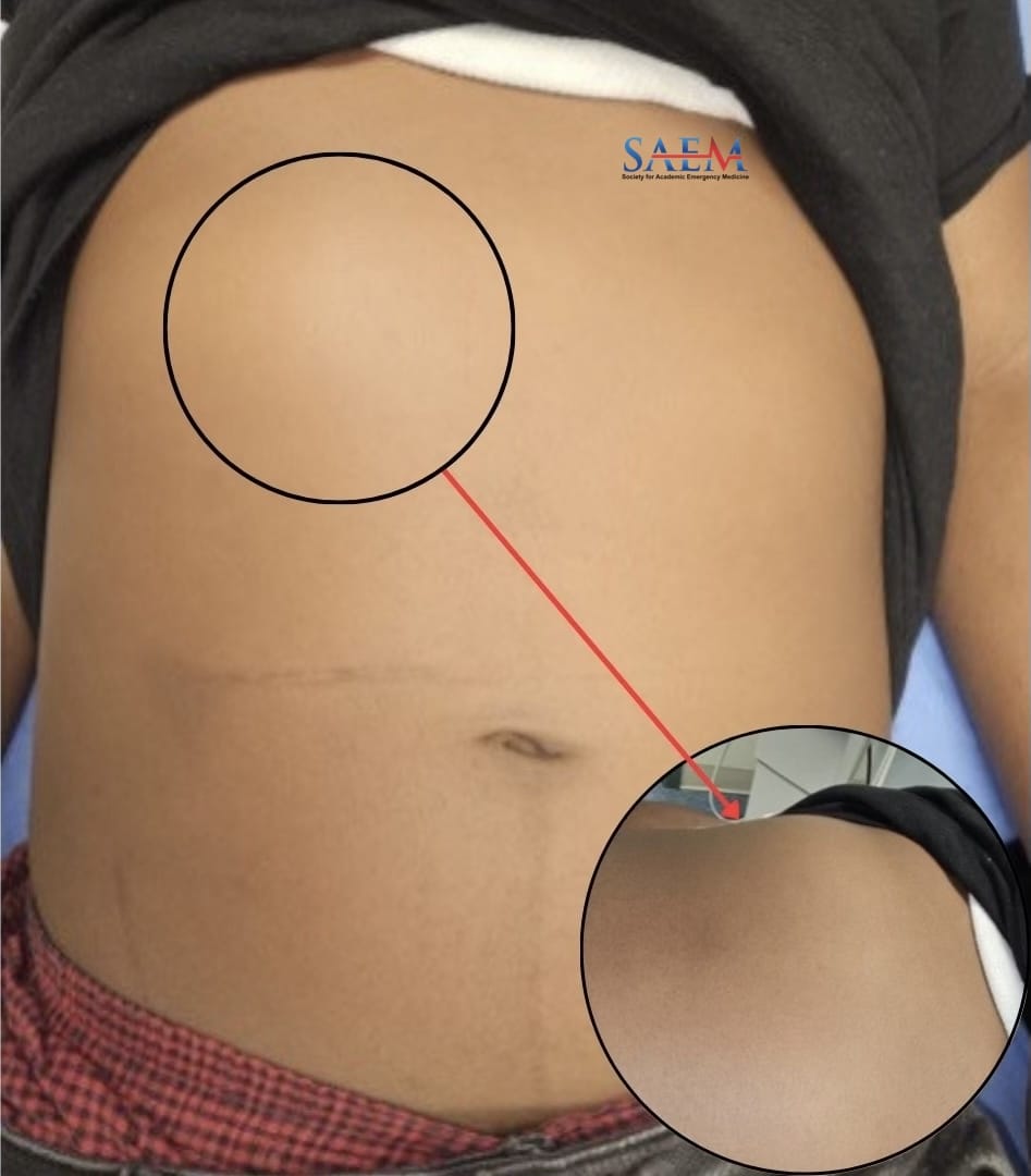
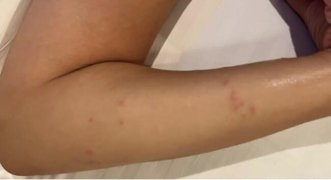
A 12-year-old male with a history of hydrocephalus status post ventriculoperitoneal (VP) shunt placement presented with an abdominal “knot.” The patient’s mother noticed the knot two days ago, on the right anterolateral thorax, which has steadily been increasing in size. The patient had no known trauma to the area or had been bitten or stung by any insect. He has otherwise been complaining of a headache, generalized, without positional changes, improved with home acetaminophen, ice pack, and rest. There were otherwise no associated vision changes, nausea, vomiting, mental status changes, or fever.

Vitals: T-36.2°C; HR 74 bpm; BP 144/75 mm Hg; RR 20; O2 Sat 96% RA

General: Well-appearing teenager in NAD.

HEENT: NC/AT. PERRL approximately 2-3 mm bilaterally. EOMI.

Neck: Supple, no meningismus.

Chest Wall: Induration to the right anterolateral thorax 5 cm x 4 cm without erythema, fluctuance, or drainage, non-tender to palpation.

Neurological: Alert. No focal neurological deficit observed.

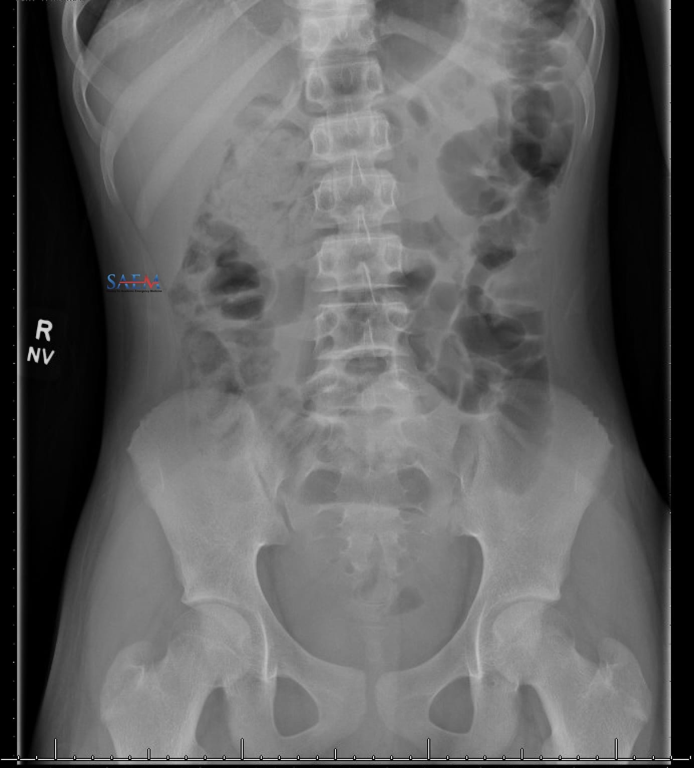
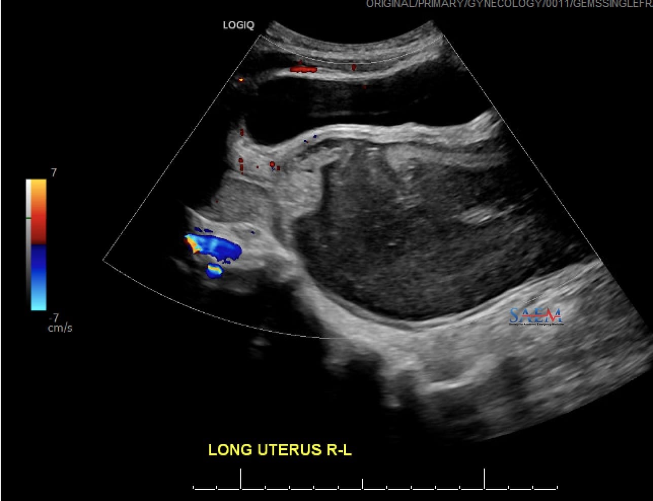
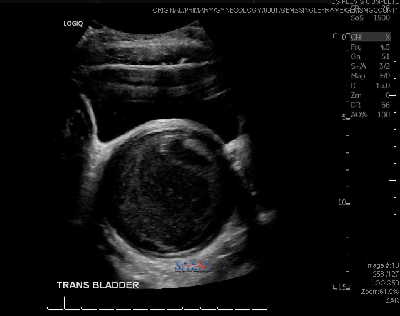
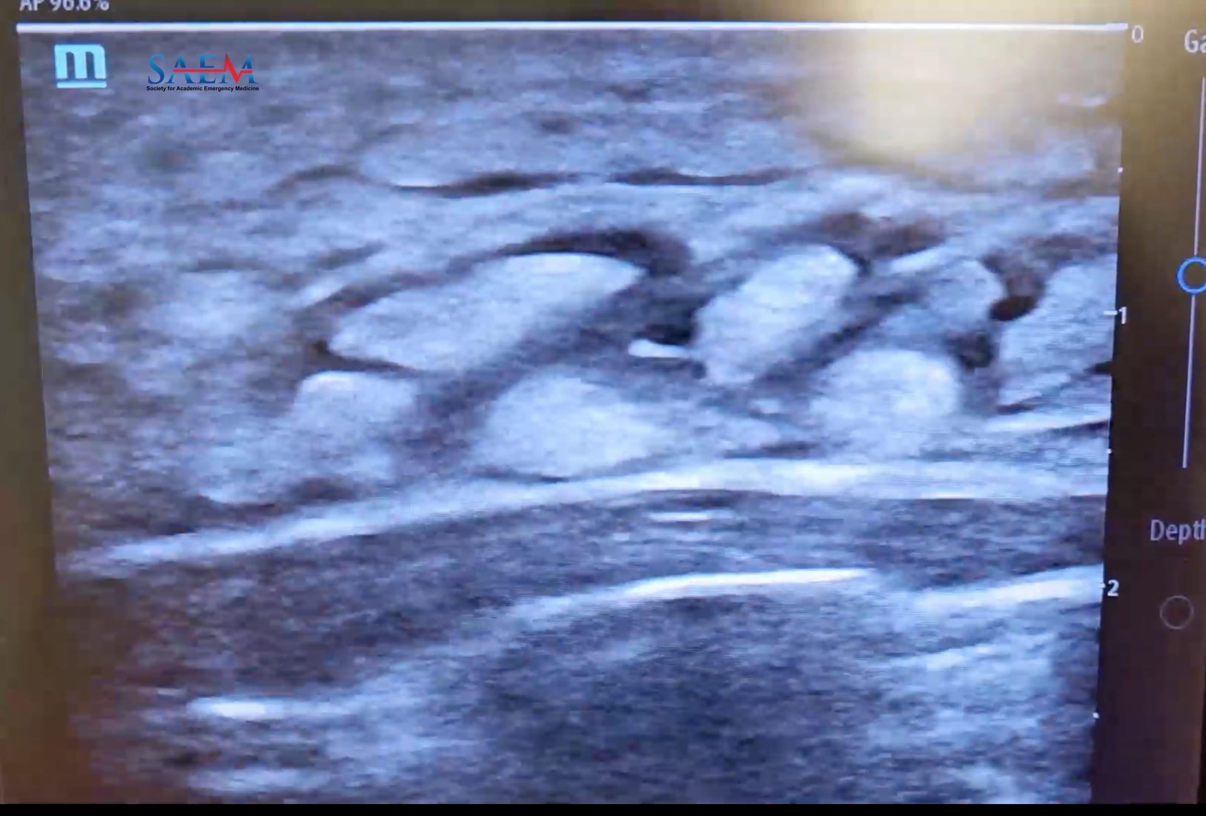
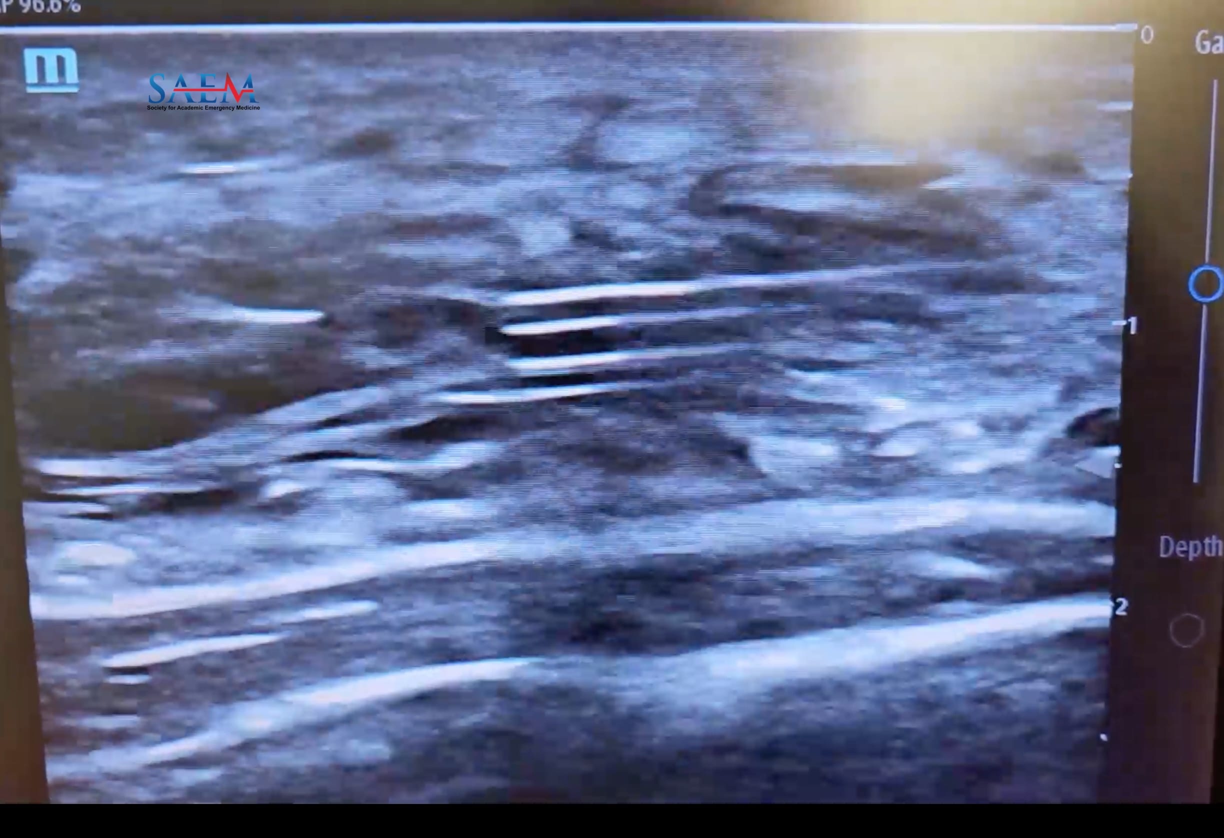
The cause of the knot is subcutaneous cerebrospinal fluid from a shunt malfunction. The ultrasound images show characteristic “cobblestoning,” indicating fluid in the subcutaneous tissue, around a linear hyperechoic object, the catheter of the VP shunt. On the plain film imaging, a disconnect was found between the thoracic and abdominal portions of the VP shunt. Up to 80% of patients with VP shunts will have experienced a shunt malfunction after 12 years, according to one study, with fractured tubing causing shunt failure in around 15% of all cases (1).

Nausea, vomiting, headache, irritability, or decreased mental status are common but nonspecific findings in shunt malfunction. Pediatric patients may present with other signs such as bulging fontanelles, increasing head circumference, or feeding and behavioral changes. An increase in the interval ventricular size can be seen in neuroimaging but can be absent in as many as 20% of patients (2). If there is a high degree of clinical suspicion for shunt malfunction, normal or unchanged neuroimaging should not preclude neurosurgical consultation.

Take-Home Points

  • In the United States, mechanical causes of VP shunt malfunction are the most common presentation, such as catheter obstruction, fracture along the clavicle or ribs, degradation of tubing, and migration of the distal catheter due to changes in height or weight.
  • Rarely, patients can develop an accumulation of CSF at the distal catheter of the VP shunt due to migration into the abdominal wall forming an abdominal pseudocyst.
  • In patients with VP shunts, abdominal complications should be considered as a sign of shunt malfunction.
  • Consider pertinent physical exam findings and POCUS to confirm the diagnosis of shunt malfunction at the distal catheter.

  • Sainte-Rose C, Piatt JH, Renier D, Pierre-Kahn A, Hirsch JF, Hoffman HJ, Humphreys RP, Hendrick EB. Mechanical complications in shunts. Pediatr Neurosurg. 1991-1992;17(1):2-9. doi: 10.1159/000120557. PMID: 1811706.

  • Reynolds RA, Ahluwalia R, Krishnan V, Kelly KA, Lee J, Waldrop RP, Guidry B, Hengartner AC, McCroskey J, Arynchyna A, Staulcup S, Chen H, Hankinson TC, Rocque BG, Shannon CN, Naftel R. Risk factors for unchanged ventricles during pediatric shunt malfunction. J Neurosurg Pediatr. 2021 Sep 24;28(6):703-709. doi: 10.3171/2021.6.PEDS2125. PMID: 34560626.

By |2025-02-19T12:57:12-08:00Feb 21, 2025|Neurology, Pediatrics, SAEM Clinical Images, Ultrasound|

ACMT Toxicology Visual Pearl: Hiss-teria Averted

snake

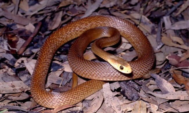
What is the predominant clinical effect of envenomation by this snake?

  1. Acute hepatic failure leading to coagulopathy
  2. Direct cardiotoxicity leading to arrhythmias
  3. Profound neuromuscular paralysis
  4. Rapid onset of shock and multisystem organ failure

[Image courtesy of iStock. ID: 1311554579]

(more…)

SAEM Clinical Images Series: A Curious Case of Anisocoria

anisicoria

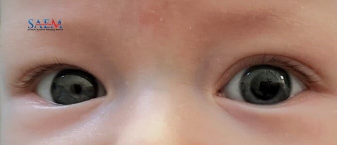
A 3-month-old male with no past medical history was brought to the emergency department for evaluation of newly asymmetric pupils. The infant appeared to be asymptomatic per parents, without any behavior changes or associated symptoms noted. The patient’s mother noticed her son’s left pupil was dilated and unresponsive to light the morning of presentation. The father had applied a prescription antiperspirant containing glycopyrronium to his axillae the previous evening but denied any known exposure to the infant.

Vitals: BP 85/66; HR 143; RR 42; SpO2 100%; T 98.3°F

Constitutional: No distress, well appearing.

HENT: Left pupil fixed and dilated to 7 mm in the light and the dark; right pupil 2 mm and reactive in the light, 5 mm in the dark. EOM intact bilaterally. No stigmata of trauma. Normal TMs bilaterally.

Neck: Normal range of motion.

Cardiovascular: Normal rate, regular rhythm and normal heart sounds.

Pulmonary: Breath sounds normal, no respiratory distress.

Abdominal: Soft, nontender, nondistended.

Neurological: Alert. Moving all 4 extremities spontaneously. Normal muscle tone. Normal suck and Moro reflexes.

Skin: Normal. No piloerection or sweating. No bruising or lesions.

No labs drawn. Head CT was obtained, which showed no acute intracranial pathology.

Ophthalmology consultation was sought, and an ophthalmologic exam demonstrated unremarkable slit lamp and fundal exams, with no afferent pupillary defect by reverse. The patient’s anisocoria was ultimately attributed to inadvertent glycopyrronium exposure from his father’s prescription antiperspirant, Qbrexza. The patient’s father later noted that he cradled the patient against his chest after applying the antiperspirant, and was not wearing a shirt at the time

Pilocarpine, a cholinergic antagonist that stimulates pupillary constriction, can be used to test mydriatic pupils. Pilocarpine drops will not reverse pharmacologically-induced anisocoria (1). Conversely, it will correct mydriasis caused by tonic pupil or third nerve palsy (2). In our patient’s case, pilocarpine administration did not result in pupillary constriction, supporting the diagnosis of drug-induced anisocoria.

Take-Home Points

  • Evaluation of acute anisocoria in the pediatric population can be challenging due to its wide range of potential etiologies including traumatic, neurologic, inflammatory, and pharmacologic causes. Though most commonly physiologic, anisocoria may represent a pediatric emergency due to the potential for underlying trauma or neurovascular compromise and thus a thorough neurologic exam and history is crucial (1, 20).

  • Inadvertent exposure to drugs such as glycopyrronium, a topical antiperspirant with anticholinergic properties, has been implicated in the pathogenesis of anisocoria in both adult and pediatric patients via inhibition of acetylcholine at the pupillary sphincter muscle (3-13). Other documented pharmacological causes of anisocoria include nebulized ipratropium bromide and scopolamine (14-19).

  • EM Clinicians should consider exposure-related anisocoria in the differential diagnosis of infant patients with acutely asymmetric pupils. In the absence of concerning neurologic findings, identification of potential drug exposures may help to minimize unnecessary testing and radiation exposure, sparing certain patients from time-intensive and costly interventions.

  • 1. Falardeau J. Anisocoria. Int Ophthalmol Clin. 2019;59(3):125-39.

  • 2. Payne WN, Blair K, Barrett MJ. Anisocoria. StatPearls. Treasure Island (FL): StatPearls Publishing Copyright © 2023, StatPearls Publishing LLC.; 2023.

  • 3. Pecha JD, Yen KG, Moisiuc A, et al. Anisocoria secondary to antiperspirant wipes in a pediatric population: a case series. J aapos. 2022;26(1):42-3.

  • 4. Chabicovsky M, Winkler S, Soeberdt M, et al. Pharmacology, toxicology and clinical safety of glycopyrrolate. Toxicol Appl Pharmacol. 2019;370:154-69.

  • 5. Coleman MJ, Tomsak RL. A 15-year-old girl with variable anisocoria. Digit J Ophthalmol. 2014;20(1):13-4.

  • 6. Micieli R, Micieli JA. Dilated Pupil in a Patient With Hyperhidrosis. JAMA. 2019;322(3):264-5.

  • 7. Radotra A, Baneke A, Paul B. Mydriasis secondary to use of glycopyrrolate cream. Br J Hosp Med (Lond). 2019;80(12):736.

  • 8. Pashaei-Marandi A, Assam JH, Arnold A, et al. Reversible anisocoria due to inadvertent ocular exposure to topical anticholinergic treatment for primary axillary hyperhidrosis. Can J Ophthalmol. 2019;54(6):e300-e2.

  • 9. Siscos SM, Figenshau K, Rajpara A. Use of gloves when applying topical glycopyrronium for treatment of primary axillary hyperhidrosis. J Am Acad Dermatol. 2020;83(4):e275.

  • 10. Kaufman AR, Gulati S, Curnyn KM. Pharmacologic anisocoria secondary to topical glycopyrronium for axillary hyperhidrosis: an emerging clinical presentation. Can J Ophthalmol. 2020;55(5):464.

  • 11. Al-Holou SN, Lipsky SN, Wasserman BN. Don’t Sweat the Blown Pupil: Anisocoria in Patients Using Qbrexza. Ophthalmology. 2020;127(10):1381.

  • 12. Kaufman AR, Gulati S, Pula JH, et al. Pharmacologic Mydriasis Secondary to Topical Glycopyrronium Tosylate Cloths: Clinical Characterization From a Multicenter Analysis. J Neuroophthalmol. 2022;42(4):530-4.

  • 13. Sandhu M, Eisenstein K. Mydriasis and anisocoria in a pediatric hyperhidrosis patient with interesting findings in the family cat. Pediatr Dermatol. 2023;40(1):210-1.

  • 14. Derinoz-Guleryuz O, Fidanci İ, Men-Atmaca Y. Nebulized Ipratropium Bromide-induced Anisocoria: Why Is Anisocoria Observed?. Iran J Allergy Asthma Immunol. 2021;20(1):125-128.

  • 15. Kokulu K, Öner H, Özen C, Eroğlu SE, Altunok İ, Akça HŞ. Pharmacologic anisocoria due to nebulized ipratropium bromide: A diagnostic challenge. Am J Emerg Med. 2019;37(6):1217.e3-1217.e4.

  • 16. Pejic R, Klaric B. Transient anisocoria in a patient treated with nebulized ipratropium bromide. Am J Ophthalmol Case Rep. 2017;7:11-13. Published 2017 Apr 12.

  • 17. Thiele EA, Riviello JJ. Scopolamine patchinduced unilateral mydriasis. Pediatrics. 1995;96(3 Pt 1):525.

  • 18. Rodor F, Cottin C, Jouglard J. Transdermal scopolamine and mydriasis. Therapie. 1989;44(6):447-448.

  • 19. Rubin MM, Sadoff RS, Cozzi GM. Unilateral mydriasis caused by transdermal scopolamine. Oral Surg Oral Med Oral Pathol. 1990;70(5):569-570.

  • 20. Gross JR, McClelland CM, Lee MS. An approach to anisocoria. Curr Opin Ophthalmol. 2016;27(6):486-492.

SAEM Clinical Images Series: Pediatric Forehead Swelling

puffy

A 12-year-old male with a history of autism spectrum disorder and chronic sinusitis presented for forehead swelling. His mother reported that she noticed progressive forehead swelling for about one month. She had followed up with the patient’s pediatrician and ENT and was given oral cephalexin and fluticasone nasal spray which did not make any changes in his symptoms. The patient denied any fevers or headaches.

Vitals: Temp 97.4°F; BP 100/58; HR 90; RR 18; SpO2 98%.

General: Patient is comfortable appearing, in no acute distress.

ENT: 3×3 cm area of fluctuance centrally located over the forehead with no drainage or surrounding erythema that is minimally tender to palpation. No nasal drainage.

Neuro: Intact with no deficits.

WBC: 14.35

ESR: 23 mm/h

CRP: 0.74 mg/dL

CT demonstrates osteomyelitis of the frontal bone with osseous destruction with a 5 cm bifrontal complex loculated anterior epidural abscess as well as a 3 cm midline frontal subgaleal extracranial scalp abscess.

Findings are consistent with Pott’s Puffy Tumor.

Take-Home Points

  • Pott’s puffy tumor is a rare, life-threatening complication of frontal sinusitis characterized by osteomyelitis of the frontal bone with associated subperiosteal abscess causing swelling and edema over the forehead and scalp. It can be found in all age groups but is most common in adolescents.
  • MRI brain with and without contrast is the preferred imaging modality due to increased sensitivity to detect early intracranial and osseous abnormalities.
  • Treatment is typically surgical intervention with at least 6 weeks of intravenous antibiotics. The infection is typically polymicrobial warranting gram-positive, gram-negative, and anaerobic antibiotic coverage.

  • Sharma P, Sharma S, Gupta N, Kochar P, Kumar Y. Pott puffy tumor. Proc (Bayl Univ Med Cent). 2017 Apr;30(2):179-181. doi: 10.1080/08998280.2017.11929575. PMID: 28405074; PMCID: PMC5349820.
  • Masterson L, Leong P. Pott’s puffy tumour: a forgotten complication of frontal sinus disease. Oral Maxillofac Surg. 2009 Jun;13(2):115-7. doi: 10.1007/s10006-009-0155-7. PMID: 19352731.

SAEM Clinical Images Series: Rectal Bulge

rectal bulge

A 13-month-old, full-term male presented due to intermittent emesis over a 3-week period. He and his parents had COVID one week prior to presentation. He had multiple episodes of non-bloody, non-bilious vomit the day before and the day of presentation. Parents noted he had been listless and unable to tolerate food. The mother was also concerned that he was straining to have bowel movements and that a mass was coming out of his bottom on the ride to the hospital. Parents reported decreased activity, decreased appetite, and decreased urine output. He was born via cesarean section due to breech presentation but had an otherwise uncomplicated prenatal history.

Constitutional: Fatigued.

Gastrointestinal: Diffuse abdominal tenderness. Reducible rectal bulge.

Skin: Pale.

Anion Gap: 19

COVID-19: Positive

WBC: 11.9

Limited Abdominal Ultrasound: A large intussusception is noted, which appears to extend at least to the descending/sigmoid colon.

XR Abdomen: Few prominent, featureless bowel loops with air-fluid levels. No gastric distention.

Air or hydrostatic enemas have a 70-85% success rate in current literature. These are often done under either fluoroscopic or ultrasound guidance. A delayed repeat enema can be done in cases where the initial enema resolved some of the intussusception. If the initial measures are unsuccessful, the patient is unstable, or the patient is exhibiting signs of peritonitis or bowel perforation, surgical management is the next step. This can either be done laparoscopically or open. In this patient’s case, an air enema was attempted but he ultimately required surgery. The surgery was laparoscopic, and he was discharged the same day.

Take-Home Points

  • Consider intussusception in any child with a URI (including COVID-19) and a rectal bulge.
  • Although this patient had a formal ultrasound, POCUS can be a useful tool in the ED to identify and expedite intussusception treatment. The classic “bullseye sign” was seen on this patient’s ultrasound.

  • Mandeville K, Chien M, Willyerd FA, Mandell G, Hostetler MA, Bulloch B. Intussusception: clinical presentations and imaging characteristics. Pediatr Emerg Care. 2012 Sep;28(9):842-4. doi: 10.1097/PEC.0b013e318267a75e. PMID: 22929138.
  • Siafakas C, Vottler TP, Andersen JM. Rectal prolapse in pediatrics. Clin Pediatr (Phila). 1999 Feb;38(2):63-72. doi: 10.1177/000992289903800201. PMID: 10047938.

SAEM Clinical Images Series: Wilma, Take a Look at This!

A 2-year, 11-month-old female with a history of constipation was brought to the ED by her mother for abdominal pain. The mother noticed that the patient’s abdomen had been enlarging for months. When they visited the pediatrician several months ago, the pediatrician also noticed a mildly enlarged abdomen but the patient was asymptomatic at that time. She was well during the interval until more recently, the patient began to complain of persistent abdominal pain and would point to the epigastric area. The patient had two episodes of unprovoked, non-bloody, non-bilious vomiting the morning prior to the ED visit. The patient had been tolerating oral intake well, passing adequate urine, having normal bowel movements, and behaving at baseline. No associated fever, diarrhea, bloody stool, dysuria, hematuria, or weight loss.

[/fusion_text]

Vitals: Temp 97.9 °F (36.6 °C); BP 103/68; Pulse 121; RR 26; SpO2 99% on room air

Constitutional: Active, well-developed, and in no distress.

HEENT: Normocephalic and atraumatic. No scleral icterus. TMs intact, no erythema. No rhinorrhea, no erythema. Moist mucous membranes, oropharynx is clear, no exudates or erythema.

Cardiovascular: Normal rate, regular rhythm and normal heart sounds.

Pulmonary: Breath sounds normal. No wheezing, no stridor, no decreased breath sounds. Normal effort, no acute respiratory distress.

Abdomen: Protuberant, distended abdomen with mild generalized tenderness to palpation. Rigid mass palpated in the upper right quadrant. Normal bowel sounds are heard.

Genitourinary: Normal anatomy. No hernias visualized, no erythema.

Skin: No jaundice or rashes visualized.

Neurological: Awake and alert. No focal deficits present.

CBC: No leukocytosis, leukopenia, anemia, or thrombocytopenia.

CMP: Electrolytes, kidney, and liver function tests were within normal limits.

The most common pediatric renal malignancy is a Wilms tumor, also known as nephroblastoma. It is an embryonal tumor due to disrupted nephrogenesis. It affects approximately 1 in 10,000 children with the median age of onset being 3.5 years (1). The most common chief complaint is abdominal pain, as in this case.

Here a large homogenous mass initially appears to be projecting from the liver, but it can also be seen protruding out of the right kidney. Pediatric abdominal organs commonly overlap so it is essential to note the origination of a mass, primarily for surgical planning. If ultrasound imaging is equivocal, CT is the next best step in differentiating the mass origination. Here, a 12 cm x 9.5 cm x 9 cm mass was noted to originate from the right kidney. If the mass becomes big enough, patients can present with vomiting due to the direct compression of the alimentary tract, such as in this case. Other presenting signs and symptoms may be fever, hypertension, anemia, hematuria, or dysuria (2).

In the US, the National Wilms Tumor Study Group recommends primary nephrectomy followed by a chemotherapy regimen that is tailored to the individual patient and tumor staging. With modern multidisciplinary management, curative therapy is achievable in approximately 90% of affected patients (2). This patient had a successful nephrectomy performed by general surgery and initiated chemotherapy on the medical floor. The patient was eventually discharged home with pediatric oncology follow-up.

Take-Home Points

  • Think of pediatric malignancy if the patient presents with chronic abdominal distention and pain.
  • Pediatric abdominal structures commonly overlap. Knowing the origination of an abdominal mass is essential for surgical planning. If ultrasound is equivocal, CT imaging is the next best step.
  • The definitive management of a Wilms tumor is a multidisciplinary approach, with primary nephrectomy followed by a tailored chemotherapy regimen as the gold-standard treatment in the US.

  1. Spreafico F, Fernandez CV, Brok J, Nakata K, Vujanic G, Geller JI, Gessler M, Maschietto M, Behjati S, Polanco A, Paintsil V, Luna-Fineman S, Pritchard-Jones K. Wilms tumour. Nat Rev Dis Primers. 2021 Oct 14;7(1):75. doi: 10.1038/s41572-021-00308-8. PMID: 34650095.
  2. Sonn G, Shortliffe LM. Management of Wilms tumor: current standard of care. Nat Clin Pract Urol. 2008 Oct;5(10):551-60. doi: 10.1038/ncpuro1218. PMID: 18836464.
  3. Leslie SW, Sajjad H, Murphy PB. Wilms Tumor. 2023 May 30. In: StatPearls [Internet]. Treasure Island (FL): StatPearls Publishing; 2023 Jan–. PMID: 28723033.

[/fusion_builder_column][/fusion_builder_row][/fusion_builder_container]

Go to Top