SplintER Series: Hip, Hip, Hooray!

A 67-year-old male with a history of bilateral total hip arthroplasties (THA) several years ago presents with left hip pain after a fall. He was walking downstairs and slipped, twisting his leg internally and with adduction and flexion of the hip to catch himself. He denies falling but felt an immediate pop in his left hip and could no longer bear weight. AP and lateral radiographs of the pelvis and left hip were obtained and are shown above (Image 1. Case courtesy of Dr Andrew Taylor, Radiopaedia.org, rID: 67457).   (more…)

Free Comprehensive Curriculum: Climate Change and Emergency Medicine

During the COVID-19 pandemic, a few of us interested in climate change science met through the Society for Academic Emergency Medicine (SAEM), and our group slowly expanded with the virtual world. We discussed the ever-growing number of climate publications and scholarship opportunities available. Some of us did research, education, or policy work, and all of us practiced clinically.

Negative climate-related impacts that we see in the Emergency Department

We discussed how climate-related impacts negatively affected our patients, and brainstormed how we could tackle the problem now. For us in Rhode Island, Pennsylvania, Wisconsin, Colorado, and California, the climate crisis was pathology and interrupted treatment regimens, but also an opportunity to transform current care systems. At all of our hospitals, patients were brought in by ambulance with empty inhalers and non-functioning medical devices after losing electrical power. Monitors beeped from abnormal vital signs of patients impacted by extreme heat, inland and coastal flooding, or wildfires. We recognized the dangers related to place of residence and structural drivers that exacerbated existing health disparities. We agreed that open access education was the next step to action and striving for justice across our nation together.

How to start your climate change learning and advocacy journey?

More and more colleagues asked us where they could begin their own climate and emergency medicine journeys. We used our varied local and global experiences to curate content that could be used for journal clubs, medical simulation, quality improvement projects, grant applications, and other educational tracks or electives. Our goal was to provide a starting place for individuals who may not have dedicated faculty at their institutions.

Get caught up: Comprehensive 10-module curriculum

Climate change and emergency medicine 10-module curriclum

We are proud to announce a comprehensive 10-module curriculum on Climate Change and Emergency Medicine (EM) worth 56 hours of ALiEMU learning credits. Each module encompasses a broad range of reading materials and is followed by a brief quiz on ALiEMU. All of this is available for free. Get learning now.

Be a climate changemaker

We hope the material reminds all of us of what actions are needed yet: authentic partnerships, clear communication of the robust evidence that we know, inclusivity, and leadership. Like emergency medicine, climate change and health work is truly life-long learning. Yet, knowledge is only as good as its use. We look forward to years of innovative solutions that move beyond dialogue and meaningfully address some of the greatest barriers to well-being for our patients and global community.

climate change and EM ALiEMU mega badge climate changer

By |2022-12-13T14:27:20-08:00Dec 14, 2022|ALiEMU, Environmental, Medical Education|

SAEM Clinical Images Series: Finger Pain

finger pain

A 57-year-old male presents after accidentally firing his son’s expired epinephrine autoinjector containing 0.3 mg of epinephrine into his left thumb two hours prior to arrival. He endorses mild pain and numbness at the tip of his thumb. The patient had spoken to poison control who had instructed him to present to the Emergency Department if his symptoms did not improve after two hours. He denied any palpitations or other systemic symptoms.

Vitals: HR 81; BP 128/81; RR 18; SpO2 99%

Extremities: Pallor noted at the distal tip of his left thumb extending 2 to 3 cm proximally with a central 2 mm area of bruising. Capillary refill could not be elicited in the left thumb pad. Sensation was intact to light touch and motor strength was intact in the left thumb.

No lab work was performed.

Treatment modalities are primarily based on case reports and range from conservative measures to local infiltration of vasodilatory medications. [1] Conservative treatments include digital massage, warm water soaks, and local application of nitroglycerin paste. Varying degrees of success have been associated with these conservative treatments. In our case, conservative measures failed and we proceed with local infiltration of phentolamine mesylate. The dosage and volume administered vary widely in reports; however, commonly reported doses were 2 to 3.5 mg of phentolamine reconstituted with 1 to 2 ml of 2% lidocaine or normal saline. This is then injected locally at multiple locations in the affected region or as a digital block. In our case, reperfusion occurred within minutes after local infiltration with approximately 1.5 mg of phentolamine reconstituted with lidocaine. No systemic side effects were noted.

There have been no documented reports of ischemic necrosis associated with accidental digital epinephrine injection. Additionally, spontaneous resolution in the majority of cases at less than two hours has been widely reported, though in some cases symptoms may persist for hours to days. [1,2] Though phentolamine may provide rapid symptomatic relief of pallor, pain, and/or paresthesia for the patient, limited data exist on long-term clinical outcomes and the overall effect on prognosis. [3]

The recommendation to never use lidocaine with epinephrine for digital nerve blocks or local digital infiltration is propagated in medical education. However, a large retrospective review of 127 accidental digital epinephrine injection cases noted a majority had spontaneous resolution. [2] Further, large studies from surgical literature have reported that local infiltration of lidocaine with epinephrine is safe and resulted in prolonged anesthesia and reduced bleeding.

Take-Home Points

  • Spontaneous resolution of accidental digital epinephrine injection often occurs within two hours and no cases of ischemic necrosis have been reported.
  • Symptomatic relief may be achieved with local phentolamine mesylate infiltration if conservative measures have failed. Commonly reported total effective doses range from 2 to 3.5 mg in volumes of 1 to 2 ml of 2% lidocaine or normal saline.

  • McNeil C, Copeland J. Accidental digital epinephrine injection: to treat or not to treat? Can Fam Physician. 2014 Aug;60(8):726-8. PMID: 25122817; PMCID: PMC4131962.
  • Muck AE, Bebarta VS, Borys DJ, Morgan DL. Six years of epinephrine digital injections: absence of significant local or systemic effects. Ann Emerg Med. 2010 Sep;56(3):270-4. doi: 10.1016/j.annemergmed.2010.02.019. Epub 2010 Mar 26. PMID: 20346537.

By |2022-12-08T21:58:38-08:00Dec 12, 2022|SAEM Clinical Images|

SplintER Series: The Hidden Post

posterior malleolar fracture

A 23-year-old male presents to the emergency department with right ankle pain after he rolled his ankle while walking down the stairs. An ankle exam reveals ecchymosis over the posterior ankle and tenderness of the distal tibia. His neurovascular exam is intact. The radiograph above was obtained (Image 1. X-ray right ankle. Original image provided by Justine Ko, MD).


(more…)

SAEM Clinical Images Series: A Rash You Don’t Want to Miss

rash

A 54-year-old female with a past medical history of diabetes presented to the Emergency Department (ED) for evaluation of unresponsiveness. The patient was found unresponsive by her spouse, who notes she had missed several doses of insulin over the past few days. EMS notes the glucometer read ‘HIGH’ on fingerstick. The patient remains unresponsive on presentation and is unable to contribute further history.

Vital Signs: BP 148/105; HR 120; RR 24; Pulse Oximetry 98% on room air; Temperature 97.7°F

Constitutional: Patient is morbidly obese, unresponsive, and toxic-appearing.

Cardiovascular: Regular rhythm with tachycardia. No murmur.

Pulmonary: Pulmonary effort is normal. Lungs clear to auscultation bilaterally.

Abdomen: Abdomen is soft and non-distended. Unable to assess for tenderness given unresponsiveness.

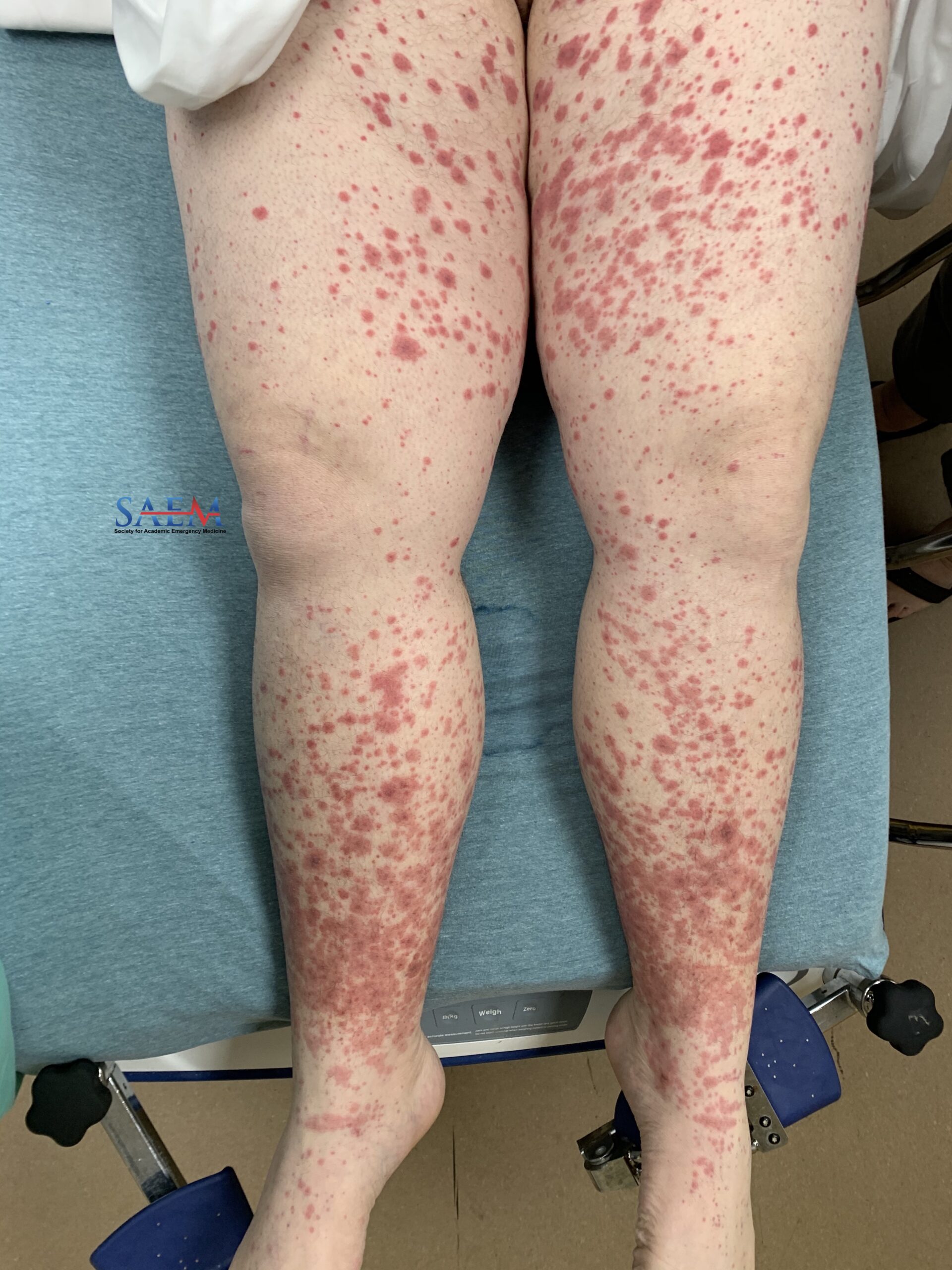
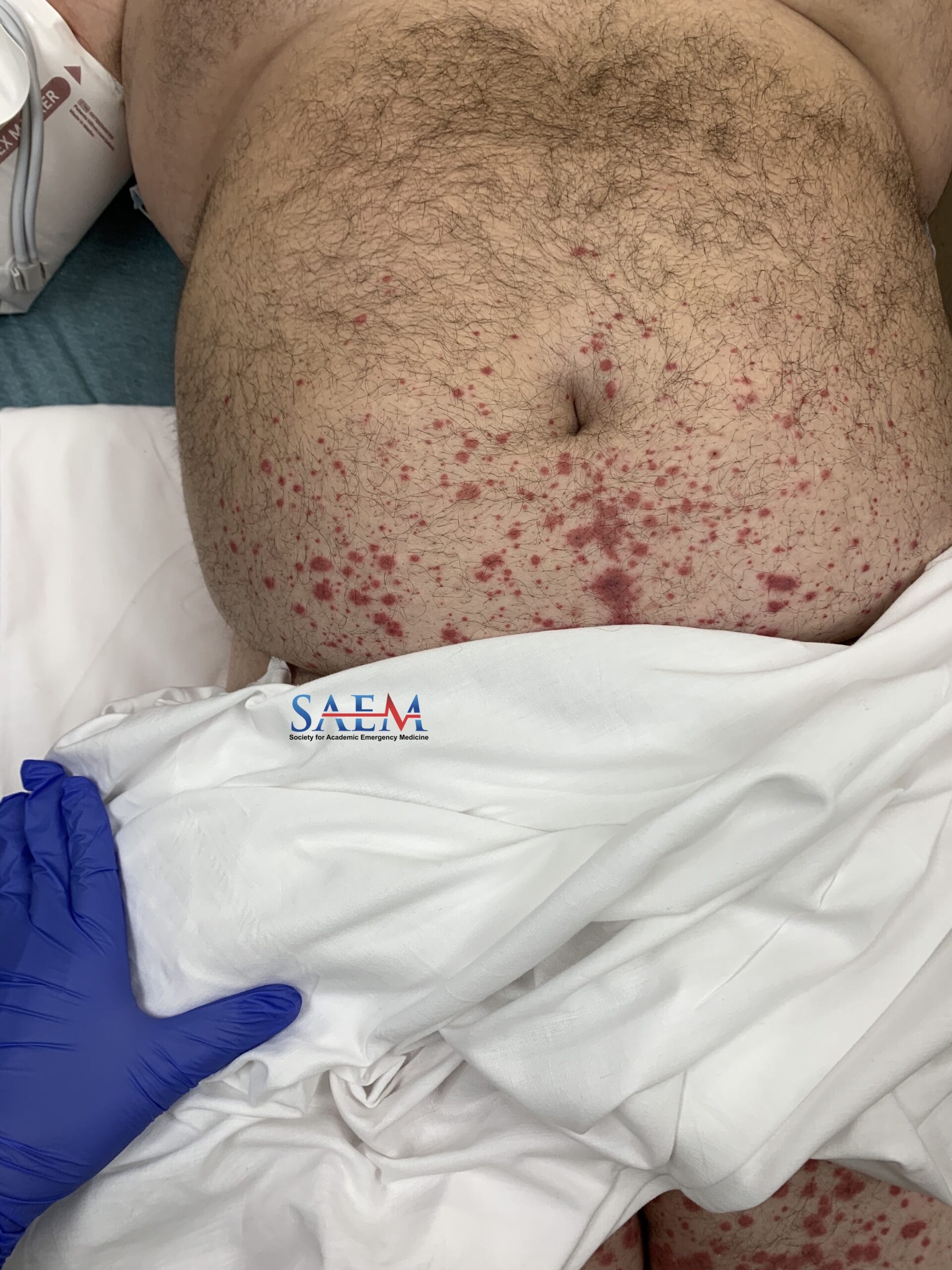
Skin: Cool, pale, mottled. Large gangrenous, draining, foul-smelling wound on proximal left thigh. There is necrotic, malodorous, black skin noted over the left lower abdomen and left upper thigh.

Neurological: Unresponsive. GCS 3.

White blood cell (WBC) count: 20.5

Comprehensive metabolic panel (CMP): K 5.8; Cr 2.06; BUN 86; Glucose >600

Venous blood gas (VBG): pH<7; lactate 3.4; bicarbonate 7

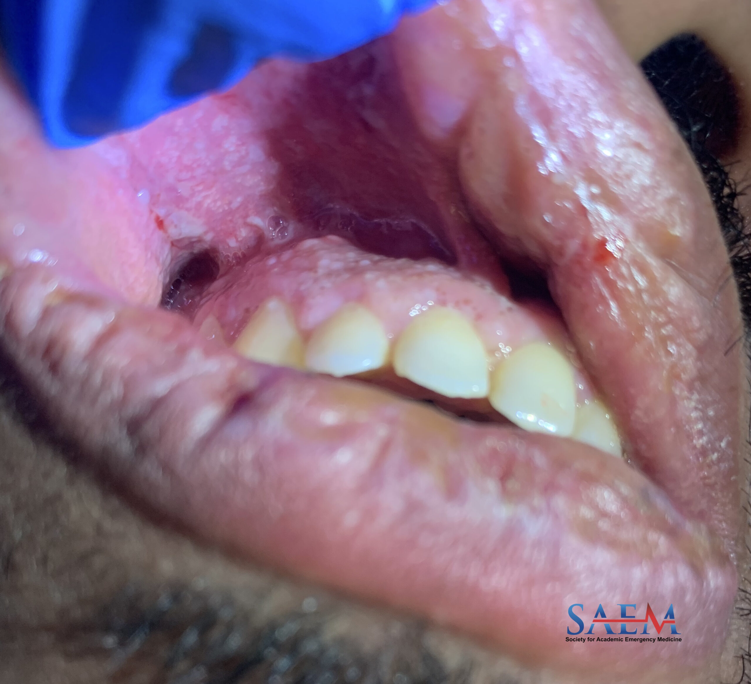
These photographs show advanced Fournier’s Gangrene, a form of necrotizing fasciitis located in the genitals, perineum, or perianal region. Rapid involvement of our surgical colleagues is crucial, as these patients will not recover without the debridement of affected tissues. Aggressive fluid resuscitation and broad-spectrum antibiotics can be initiated after a surgical consult is made.

This patient presented with impressive visual evidence of advanced disease including severe ecchymosis, but our clinical suspicion must be high as Fournier’s is rapidly progressing and carries a high mortality rate (may be upwards of 30%). Earlier symptoms are genital or perineal pain which may be associated with itching, lethargy, or fever. The biggest exam finding to keep in mind is ‘pain out of proportion to the exam’ as up to 40% of these patients may present without localized symptoms. Advanced disease, as seen in this patient, can present with crepitus and severe ecchymosis of tissue involved. This patient was also found to be in DKA, as evidenced by her laboratory findings. This case should serve as a reminder that it is vital to perform a proper skin examination in patients presenting with hyperglycemia. Ultimately in this case, the patient was intubated for airway protection and started on vasopressors for cardiovascular support in the setting of septic shock. She went into VTach arrest and was successfully defibrillated before further decompensating and becoming asystolic.

Take-Home Points

  • The first task after suspected diagnosis of Fournier’s Gangrene is a page to your surgery service for evaluation and emergent debridement in the OR (depending on your institution this may be general surgery, urology, or both). After your patient is on the path to definitive management, you can begin aggressive fluid administration and broad-spectrum antibiotics (gram-positive, gram-negative, and anaerobic coverage needed).
  • Fournier’s Gangrene is a clinical diagnosis. Imaging may assist in atypical or borderline cases, but should never result in delay of surgical evaluation and treatment. Crepitus and ecchymotic tissue are very late findings; have high clinical suspicion inpatients with signs of swelling, erythema, and pain.

  • Shyam DC, Rapsang AG. Fournier’s gangrene. Surgeon. 2013 Aug;11(4):222-32. doi: 10.1016/j.surge.2013.02.001. Epub 2013 Apr 8. PMID: 23578806.
  • Ustin JS, Malangoni MA. Necrotizing soft-tissue infections. Crit Care Med. 2011 Sep;39(9):2156-62. doi: 10.1097/CCM.0b013e31821cb246. Erratum in: Crit Care Med. 2011 Nov;39(11):2592. Dosage error in article text. PMID: 21532474.
  • Harbrecht BG, Nash NA. Necrotizing Soft Tissue Infections: A Review. Surg Infect (Larchmt). 2016 Oct;17(5):503-9. doi: 10.1089/sur.2016.049. Epub 2016 Aug 2. PMID: 27483003.
  • Singh A, Ahmed K, Aydin A, Khan MS, Dasgupta P. Fournier’s gangrene. A clinical review. Arch Ital Urol Androl. 2016 Oct 5;88(3):157-164. doi: 10.4081/aiua.2016.3.157. PMID: 27711086.
  • Sarani B, Strong M, Pascual J, Schwab CW. Necrotizing fasciitis: current concepts and review of the literature. J Am Coll Surg. 2009 Feb;208(2):279-88. doi: 10.1016/j.jamcollsurg.2008.10.032. Epub 2008 Dec 12. PMID: 19228540.
  • Tintinalli JE, Ma O, Yealy DM, Meckler GD, Stapczynski J, Cline DM, Thomas SH. eds. Tintinalli’s Emergency Medicine: A Comprehensive Study Guide, 9e. McGraw Hill; 2020. p.592- 593.

SAEM Clinical Images Series: ‘Tis Not the Season to be Wheezing

wheezing

A 2-year-old male with a history of solitary kidney presented with greater than one month of daily coughing, wheezing, and decreased appetite. The patient was previously seen by his primary care physician after three weeks of symptoms where he was prescribed albuterol as needed for viral bronchospasm. The patient’s wheezing did not improve after two weeks of albuterol treatment so a chest x-ray was ordered. The patient’s mother denied any fevers, vomiting, diarrhea, weight changes, or night sweats.

Vitals: BP 131/60; Pulse 148; Temp 36.7 °C (98.1 °F) (Axillary); Resp 28; Wt 15.7 kg (34 lb 9.8 oz); SpO2 95%

General: Alert; well appearing

HEENT: Pupils equally reactive to light; moist mucous membranes; nares with normal mucosa without discharge

Cardiovascular: Regular rate; regular rhythm; normal S1, S2; no murmur noted; distal pulses 2+

Pulmonary: Good aeration throughout all lung fields; clear breath sounds bilaterally; prolonged expiratory phase; stridor with agitation

Abdomen: Soft; non-tender; non-distended

White blood cell (WBC) count: 56.1/uL (Blasts 58%)

Platelets: 288/uL

Uric acid: 8.3 mg/dL

LDH: 2231 iU/LD

D-Dimer: 3.22 ug/mL

Fibrinogen: 463 mg/dL

Bronchospasm, bronchiolitis, viral infection, pneumonia, foreign body aspiration, space-occupying lesion, vocal cord dysfunction, cardiac dysfunction, and acute chest in patients with sickle cell disease.

The radiograph shown demonstrates a mediastinal mass. This patient was ultimately diagnosed with T-cell acute lymphoblastic leukemia. T-ALL can present with fatigue, fevers, weight loss, easy bleeding/bruising, paleness, or a mediastinal mass. Mediastinal masses found on chest x-ray require further evaluation to determine the diagnosis, location, and treatment. If malignancy is suspected, an oncology referral and bone marrow sample will be necessary.

Take-Home Points

  • In patients with first-time wheezing that does not improve with bronchodilator therapy, consider alternative diagnoses and further evaluation.
  • A mediastinal mass is found at the time of diagnosis in 10% to 15% of children with acute lymphoblastic leukemia.

  • Steuber, P (2021). Overview of common presenting signs and symptoms of childhood cancer.UpToDate. Retrieved January 2, 2021.2.
  • Juanpere, S., Cañete, N., Ortuño, P., Martínez, S., Sanchez, G., & Bernado, L. (2013). A diagnostic approach to the mediastinal masses. Insights into imaging, 4(1), 29–52.https://doi.org/10.1007/s13244-012-0201-0

Trick of the Trade: Managing Epistaxis with Merocel Nasal Packing and an Angiocatheter


There are many ways to manage epistaxis. Once nasal clamping and cauterization fail, the next step is to consider using tranexamic acid (TXA) and performing nasal packing. Inflatable packing devices such as a Rhinorocket are painful to insert and do not conform well to the shape of the naris. The expandable Merocel nasal packing, a compressed, dehydrated sponge, provides a softer, alternative option, although the insertion process can be painful given its initial rigid, edged structure. We propose 2 strategic tricks to optimize your nasal packing technique using the Merocel sponge.

Trick of the Trade: Strategic expansion of the Merocel sponge

The common approach for Merocel packing involves inserting the unexpanded sponge into the nose, tilting the patient’s head back, and dripping in TXA solution to expand the sponge to tamponade the bleeding.

Trick #1: Wet the tip of the Merocel’s sharp edge to allow for a softer cushion to slide the packing more comfortably and deeper into the naris.

Trick #2: Use an angiocatheter to deliver the TXA solution directly onto the mid-portion of the packing. Commonly, the TXA solution is dripped onto the outer end, which may cause an uneven and inadequate expansion at the site where the bleeding may be occurring. Because blood also can react with the packing, it is likely the blood will expand the packing before TXA reaches the center by osmosis. Another benefit of Merocel expansion starting at the center is that it will help anchor the sponge in place. In contrast, TXA administration at the outer tip first may pull the sponge out of the naris a few millimeters.

Equipment

  • 20g or 22g angiocatheter (closed IV catheter system)
  • Tranexamic acid solution
  • A syringe
  • Merocel nasal dressing

Technique

merocel sponge nasal packing trick setup

1. Insert the angiocatheter needle into the Merocel packing about ⅓ the distance from the external end of the packing. Remove the needle, leaving the plastic angiocatheter in place.

merocel tip moisten txa trick

2. Soak the insertion tip of the nasal packing with a drop of TXA to soften it. Or apply a light coat of an antibiotic ointment or petroleum jelly to the insertion tip for lubrication. This will make it easier to advance the packing and also less painful for patients. Advance the Merocel into the affected naris just as you would a nasogastric tube. Some additional tips are in the ALiEM article about nasogastric and nasopharyngeal tube insertion.

3. Once the nasal packing is fully inserted, expand the sponge by administering TXA via the attached angiocatheter. The mid-portion of the sponge should expand first, thus preventing outward slippage of packing. Also TXA more quickly reaches the area of bleeding rather than from a more gradual osmotic effect when dripped in from the external tip.

Go to Top