Patwari Academy videos: Toxicology

What is your approach to the poisoned patient? Listen to these 4 videos by Dr. Rahul Patwari to build your foundation of knowledge in toxicology.
- Introduction
- Toxidromes
- Testing
- Decontamination



What is your approach to the poisoned patient? Listen to these 4 videos by Dr. Rahul Patwari to build your foundation of knowledge in toxicology.
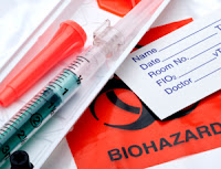 You obtain a venous blood gas (VBG) on a patient with a COPD exacerbation because you are concerned about hypercarbia. You get a value of 55 mmHg. How correlative is that compared to an arterial blood gas (ABG). There has been a lot of literature on how well the pH correlates between the ABG and VBG but what about pCO2?
A small study (n=89) from 20121 found that with a cutoff of pCO2 < 45 mmHg, the venous pCO2 is 100% sensitive in ruling out arterial hypercarbia. When the pCO2 was ≥ 45 mmHg, the VBG was less correlative.
Below is a review by Dr. Michelle Reina (EM resident at Univ of Utah) and Dr. Rob Bryant (Intermountain Medical Center in Utah) of the VBG vs ABG correlative data, along with a proposed algorithm on what to do with patients with COPD exacerbation.
Adapted from [1–5]
Go to ALiEM (PV) Cards for more resources.
Updated 1/31/13 at 2 pm PST:

As a nice segue from the Low Risk Chest Pain videos, below is a 3-part series on Demystifying the Electrocardiogram by Dr. Rahul Patwari. It takes talent to make the complex simple.
(more…)

A patient presents with an asymmetric leg with trace pitting edema in the affected leg. What is your diagnostic approach to such a patient? What is the role of D-dimer and ultrasound (U/S)? Does this match the 2012 American College of Chest Physicians (ACCP) guidelines?
The first step is to determine your patient’s pretest probability because the recommendations vary based on risk. I can tell you that many ED patients come in with a Wells score of 1-2, which places them in the “moderate pretest probability” category. There are 2 approaches you can take based on the availability of resources at your site (high-sensitivity D-dimer or U/S) and the patient’s comorbidities. Are you referring your patient for a repeat outpatient ultrasound, if warranted?Walk through various patient scenarios to see how the D-Dimer and U/S come into play.
Adapted from [1]
Go to ALiEM (PV) Cards for more resources.
Thanks to Dr. Jason West (EM resident at Jacobi/Montefiore) for this card idea and deciphering the complex recommendations from the publication.
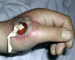
The technique for abscess drainage traditionally is incision and drainage (I&D). In August 2012, I wrote about incision and loop drainage (I&LD), which it seems has gained popularity over time with similar outcomes. This technique involves using a sterile vascular loop, which is thin and long enough to form a loose knotted loop. The video below by Dr. Rob Orman reviews the steps. But, what if you don’t have a vascular loop in the ED?

One of the most common complaints in the Emergency Department is chest pain. Is it something serious? How do I risk-stratify patients with potential acute coronary syndrome? What should I be thinking of and not missing?
Rahul goes over the low-risk chest pain patient in 2 great, short teaching videos, based partly on the 2010 AHA/ACC Guideline for ACS risk stratification.