



Announcing ALIEM Remote
We are proud to announce the ALiEM Remote series. 2020 has presented a challenging set of circumstances with an ongoing COVID pandemic, uncertainty in the workplace, and multiple educational closures at every level. With our remote series, we aim to help ameliorate these challenges by providing you with great tips and tricks for remote work, remote education, and a list of speakers ready and willing to go virtual for your next department meeting or residency conference. We want to provide everyone in emergency medicine a centralized place to find resources on how to work, learn, educate, and live remotely. [+]
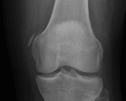
SplintER Series: Delayed pain in an injured knee
Image 1. Case courtesy of Radswiki, Radiopaedia.org A 26 year-old male presents with new medial right knee pain after twisting his knee playing soccer 3 weeks ago. His initial pain has since resolved. What is your diagnosis? What examination findings should you expect? What associated diagnoses should you assess for? What is your management in the emergency department? [+]
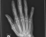
Diagnose on Sight: Post-traumatic Finger Pain
Case: A 32-year-old male with a past medical history of diabetes presents with a 1 month history of finger pain after slamming his finger in a car door. 2 weeks after the initial incident he presented to the emergency department for worsening pain and received x-rays that were negative for acute fracture. Today he presents reporting pain radiating up the hand, arm, and into the shoulder, with associated chills. His labs are significant for hyperglycemia, hyponatremia, and an elevated erythrocyte sedimentation rate and c-reactive protein. His x-ray is seen here (figure 1 image courtesy of Daniel Rogan, MD). What is [+]
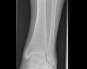
EMRad: Can’t Miss Adult Ankle and Foot Injuries
Have you ever been working at 3am and wondered, “Am I missing something? I’ll just splint and instruct the patient to follow up with their PCP in 1 week.” This is a reasonable approach, especially if you’re concerned there could be a fracture. But we can do better. Enter the “Can’t Miss” series: a series organized by body part that will help identify injuries that ideally should not be missed. This list is not meant to be a comprehensive review of each body part, but rather to highlight and improve your sensitivity for these potentially [+]
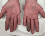
Case of a Blue, Painful Finger
A 37-year-old female presented to the emergency department for evaluation of numbness and discoloration to her left fourth finger, which had started the day before. The patient stated that she was gardening the previous day and afterward she noticed the discoloration and pain. The patient denied taking any medications. She reported recreational methamphetamine and heroin use. She denied any chest pain or difficulty breathing. She denied any history of Raynaud’s phenomenon, venous thromboembolism, or history of trauma. The patient was afebrile with normal vital signs. Physical exam revealed cyanotic discoloration to the left distal fourth finger. Sensation was intact to [+]
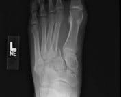
EMRad: Approach to the Traumatic Foot X-ray
Radiology teaching during medical school is variable, ranging from informal teaching to required clerkships [1]. Many of us likely received an approach to a chest x-ray, but approaches to other studies may or may not have not been taught. We can do better! Enter EM:Rad, a series aimed at providing “just in time” approaches to commonly ordered radiology studies in the emergency department. When applicable, it will provide pertinent measurements specific to management, and offer a framework for when to get an additional view, if appropriate. We recently covered the elbow, wrist, and ankle: now, the foot x-ray. [+]
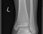
EMRad: Radiologic Approach to the Traumatic Ankle
Radiology teaching during medical school is variable, ranging from informal teaching to required clerkships [1]. Many of us likely received an approach to a chest x-ray, but approaches to other studies may or may not have not been taught. We can do better! Enter EM:Rad, a series aimed at providing “just in time” approaches to commonly ordered radiology studies in the emergency department. When applicable, it will provide pertinent measurements specific to management, and offer a framework for when to get an additional view, if appropriate. We recently covered the elbow and wrist. Now: the ankle. [+]
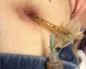
Trick of the Trade: Deflate an Undeflatable Gastrostomy Tube
A 54-year-old female with a past medical history of throat cancer presents for gastrostomy tube (G-tube) replacement. The initial G-tube was placed 3 years ago. Most recently, the patient had the G-tube changed 7 months ago. She presents to the Emergency Department because the G-tube is leaking from the tubing that is external to the skin. When you attempt to deflate the cuff, you are unsuccessful. [+]
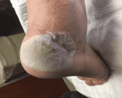
ACMT Toxicology Visual Pearls: Painful Foot
Question: A patient presents with foot pain 4 hours after contact with a chemical in the garage. What chemical exposure likely caused this injury? Ethylene Glycol Hydrofluoric Acid Petroleum Propylene Glycol Sodium Hypochlorite [+]

Ethen Ellington
Carolinas Medical Center
Charlotte, NC ,br>
Latest posts by Ethen Ellington [+]
Navigating Life After Residency: 10 Lessons I Learned in EMS Fellowship
The transition from residency to your first job or fellowship is an exciting time in any career. New opportunities for professional growth appear, but with them come a new and unique set of challenges. Transitioning from a structured clinical environment to more independent work and self-driven projects can be a difficult transition. For this reason, we wanted to share a few lessons we’ve learned. Although this advice is derived from our experience in EMS fellowship, we expect that it will apply and be helpful to other upcoming fellows and all people stepping away from residency to enter the workforce. [+]

Director of Growth, Strategic Alliance Initiative, Center for Innovation and Digital Health
Massachusetts General Hospital;
Chief Scientific Officer, Conductscience.com
Latest posts by Shuhan He, MD (see all)
- The Discharge Severity Index: Early Research on ED Readmission Risk Assessment - August 14, 2025
- Suboxone and the Emergency Physician: Get Waivered Training - August 12, 2022
- A Tale of Two Epidemics: COVID-19 and the Opioid Crisis - April 23, 2020