


ALiEM AIR Series | Vascular Module (2025)
Welcome to the AIR Vascular Module! After carefully reviewing all relevant posts in the past 12 months from the top 50 sites of the Digital Impact Factor [1], the ALiEM AIR Team is proud to present the highest quality online content related to related to HEENT emergencies in the Emergency Department. 8 blog posts met our standard of online excellence and were approved for residency training by the AIR Series Board. More specifically, we identified 3 AIR and 5 Honorable Mentions. We recommend programs give 4 hours of III credit for this module. AIR Stamp of Approval and Honorable [+]
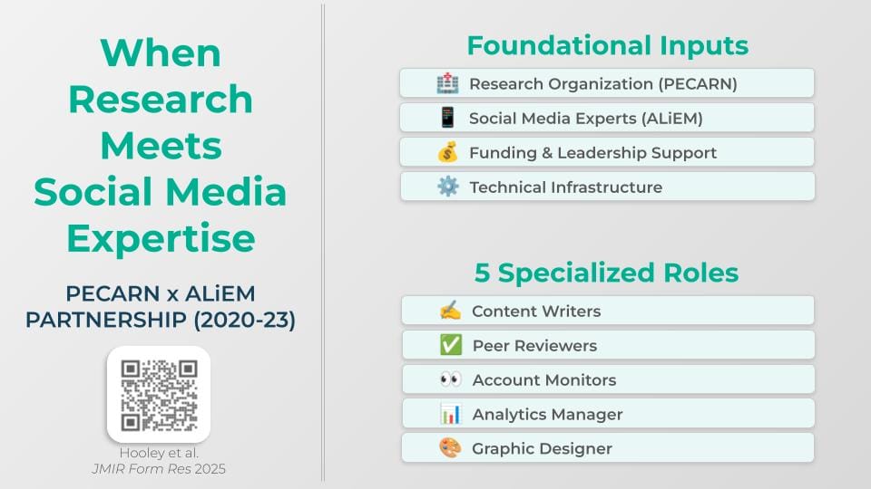
When Research Meets Social Media Expertise: Lessons from the PECARN-ALiEM Partnership
From Pipe Dream to Proven Strategy: How a 4-year partnership between PECARN and ALiEM created a replicable framework for evidence-based research dissemination Sometimes the best collaborations begin with simple questions. Following Dr. Nathan Kuppermann's grand rounds presentation in 2018, I had the opportunity to discuss an idea with him as PECARN's Steering Committee Chair: might there be untapped potential in using social media platforms like Twitter to amplify PECARN's research impact? Five years later, that initial conversation has grown into a reality with a systematic approach and measurable outcomes. Social media is not just about fads and marketing. In [+]
ACMT Toxicology Visual Pearl: Not Very A-peel-ing
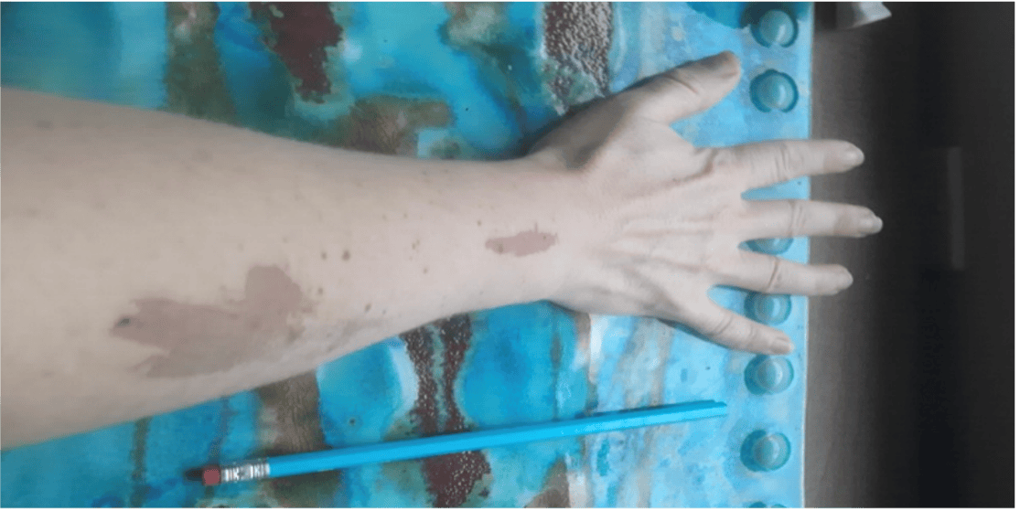
Which of the following chemicals, commonly used in chemical peels, can cause severe, gray colored skin burns upon direct contact? Acetone Hydrogen peroxide Phenol Salicylic acid [Author’s own image] [+]

Emma Lindemann, MD
Corewell Health West - Butterworth Hospital
Grand Rapids, MI
Latest posts by [+]
PEM POCUS Series: Pediatric Cardiac
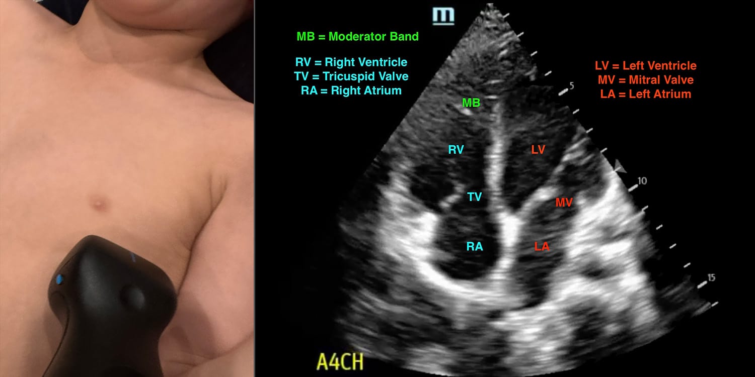
Read this tutorial on the use of point of care ultrasonography (POCUS) for pediatric cardiac evaluation. Then test your skills on the ALiEMU course page to receive your PEM POCUS badge worth 2 hours of ALiEMU course credit. Take the ALiEMU PEM POCUS: Pediatric Cardiac Module Goals List the indications and limitations of pediatric cardiac point-of-care ultrasound (POCUS) Describe the technique for performing cardiac POCUS Identify anatomical landmarks accurately on cardiac POCUS Interpret abnormal findings on cardiac POCUS Describe the basic literature behind pediatric cardiac POCUS Case Introduction: Child with respiratory distress You are in [+]
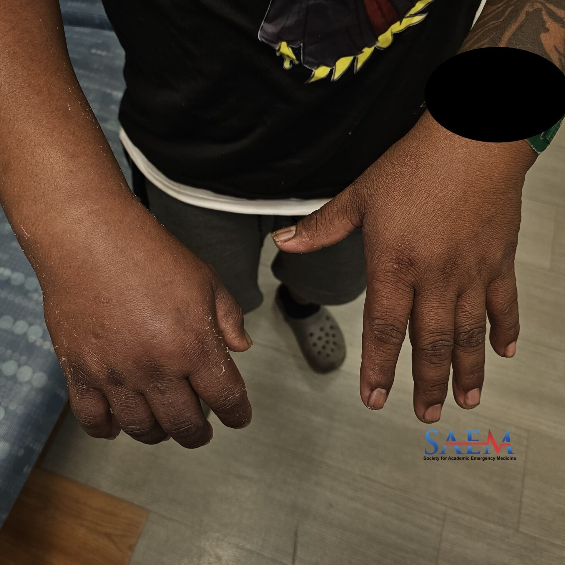
SAEM Clinical Images Series: Pain and Swelling in a Roofer’s Right Wrist
A 27-year-old male with no significant past medical history presented to the ED due to right hand pain and swelling. The patient reported that he works as a roofer and felt severe, sharp pain in his right hand immediately after using a nail gun this morning. The pain was followed by gradual swelling of the right wrist and hand. There was no loss of sensation or bleeding from the injury site. He additionally denied any injury from the nail itself. The patient was in moderate pain but hemodynamically stable while in the ED. [+]
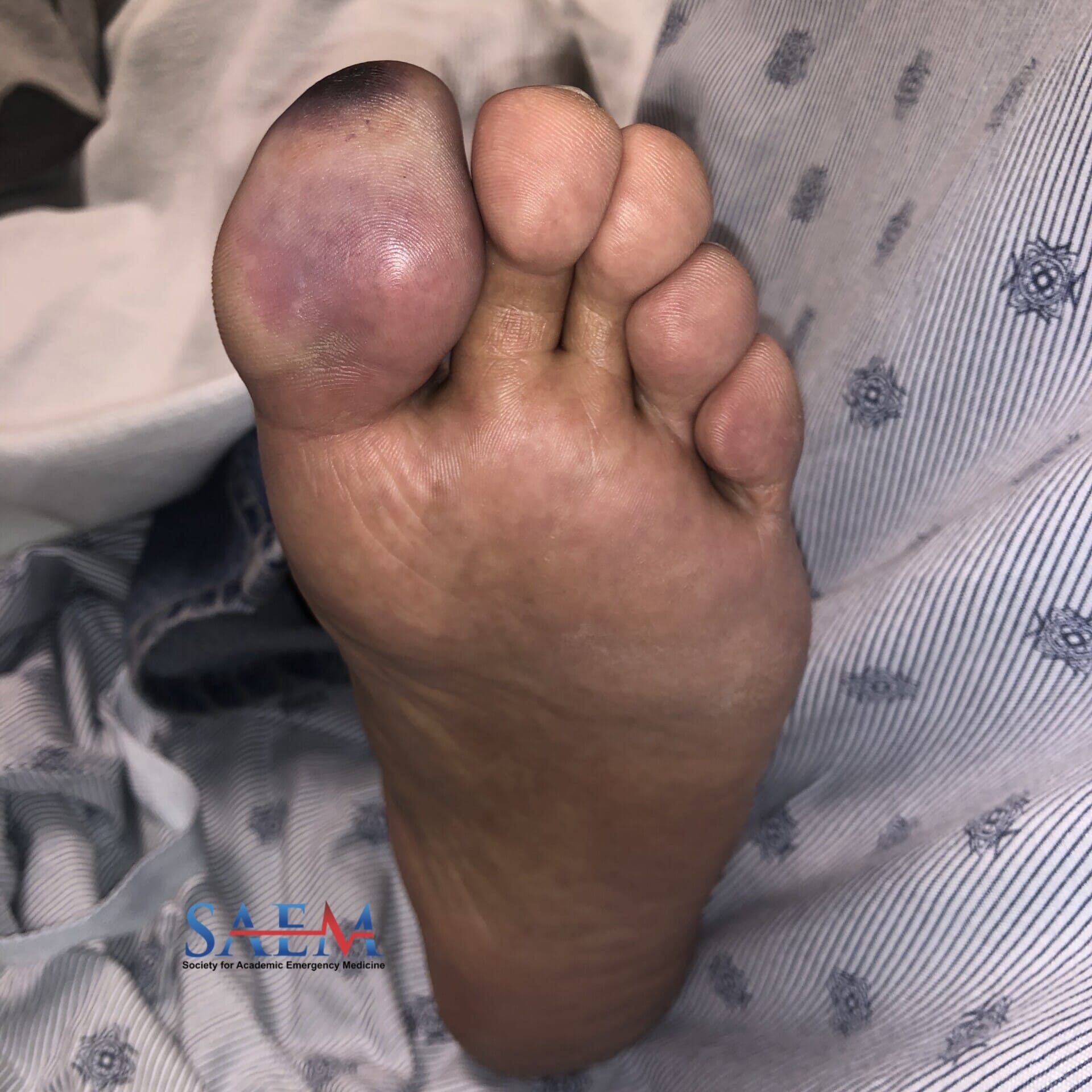
SAEM Clinical Images Series: Painful Great Toe
A 63-year-old-male with a past history of hypertension, renal failure with dialysis three times per week, and prior infective endocarditis appropriately treated with a regimen that ended four weeks ago presented with left great toe pain that started three weeks ago. The toe began swelling two weeks ago with continued pain and tenderness, becoming discolored approximately one week ago. He noted subjective fever and chills, but had no other complaints. Vitals: Heart Rate 104, BP 111/69 mmHg, Temperature oral 38.1°C, Respiratory Rate 16; SpO2: 99% on room air Skin: The plantar [+]
ALiEM AIR Series | Environmental Module (2025)
Welcome to the AIR Environmental Module! After carefully reviewing all relevant posts in the past 12 months from the top 50 sites of the Digital Impact Factor [1], the ALiEM AIR Team is proud to present the highest quality online content related to related to Environmental emergencies in the Emergency Department. 5 blog posts met our standard of online excellence and were approved for residency training by the AIR Series Board. More specifically, we identified 1 AIR and 4 Honorable Mentions. We recommend programs give 3 hours of III credit for this module. AIR Stamp of Approval and Honorable Mentions [+]
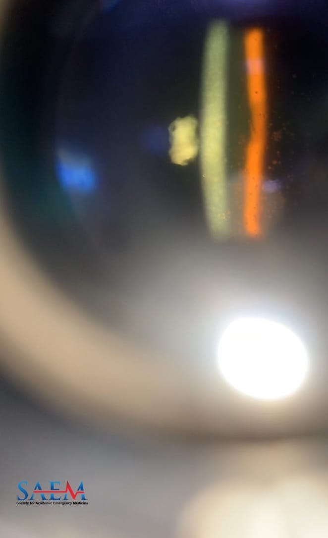
SAEM Clinical Images Series: I Cannot See My Rashes
A 37-year-old African-American transgender patient presented with progressive, bilateral painful vision loss. The symptoms began acutely in the right eye two weeks prior to presentation, eventually extending to the left eye. Symptoms were worse in the right eye and included headache, blurry vision, photophobia, and pain with eye movement. Additionally, the patient reported the appearance of a diffuse, generalized, non-pruritic, non-tender rash of unknown duration or timeline. The rash was hyperpigmented and located on the trunk, face, genitalia, palms, and soles. The patient denied any recent trauma, using eye drops, wearing glasses, recent new detergents, [+]
SAEM Clinical Images Series: I’m Plugged Up
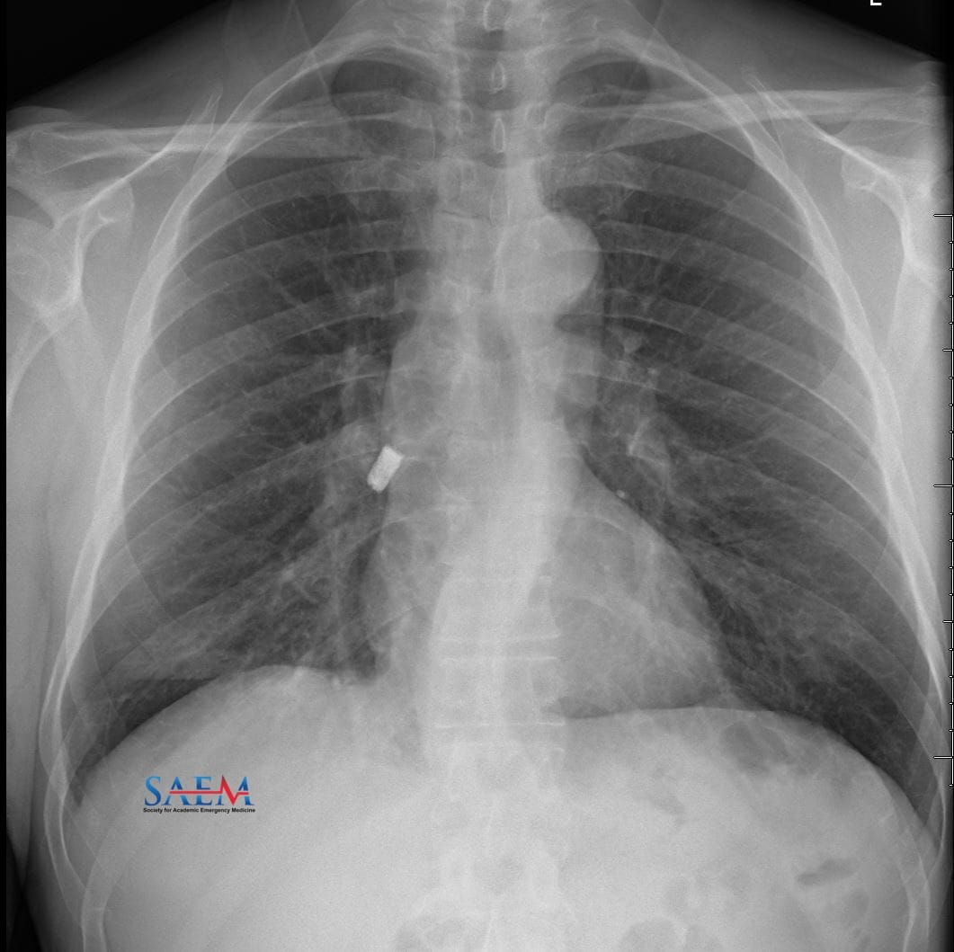
A 56-year-old male with a history of crack cocaine and opiate drug use presented to the Emergency Department (ED) with concerns about a persistent dry cough for seven days. The cough started after smoking cocaine from a homemade glass pipe. The day before arrival, he developed sharp chest pain with coughing associated with shortness of breath and a sore throat. The patient denied fevers, chills, or shortness of breath. He denied hemoptysis, nausea, vomiting, and diarrhea. A chest x-ray was obtained with the findings seen below. Vitals: Temp: 36.6°C; HR: 78 [+]
ACMT Toxicology Visual Pearl: Turning Blue
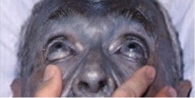
What commercially available product can cause blue-grey discoloration of the skin and conjunctiva with long term use? Benzocaine, 20% oral gel Colloidal silver, 30 ppm liquid solution Ferrous sulfate, 325 mg tablet Methylene blue, 1% oral solution [Image from Herbert L. Fred, MD and Hendrik A. van Dijk via Wikimedia Commons] [+]

Brandon Friedman, MD

Director of Growth, Strategic Alliance Initiative, Center for Innovation and Digital Health
Massachusetts General Hospital;
Chief Scientific Officer, Conductscience.com
Latest posts by Shuhan He, MD (see all)
- The Discharge Severity Index: Early Research on ED Readmission Risk Assessment - August 14, 2025
- Suboxone and the Emergency Physician: Get Waivered Training - August 12, 2022
- A Tale of Two Epidemics: COVID-19 and the Opioid Crisis - April 23, 2020