


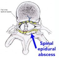
High risk back pain: Spinal Epidural Abscess
Spinal epidural abscess (SEA) is a rare but potentially catastrophic cause of back pain. Classically these patients are described as having back pain, fever, and clear neurologic deficits. In reality, patients often present with less obvious symptoms which often leads to a delay in diagnosis. Missed cases of SEA are a source of significant risk to both the patient and the provider. To improve outcomes and minimize risk, providers must identify and promptly evaluate patients who are at increased risk of developing a SEA. [+]
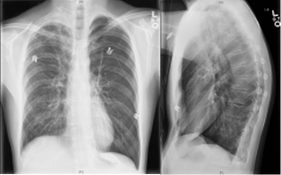
Diagnose on Sight: “I feel like I am having a heart attack”
Case: A 18-year-old male with no medical history presents to the emergency department (ED) complaining of “feeling like I am having a heart attack” which started suddenly 1 hour ago. The patient ate from a food truck the night before and developed several episodes of forceful vomiting prior to arrival in the ED. What finding in this supine chest radiograph aids in the diagnosis? Click on image for a larger view. [+]

Vomiting in Kids After Head Trauma: To CT or Not to CT?
A 6-year-old male is brought to the emergency department (ED) after falling from the monkey bars at a local playground. Physical examination reveals no scalp hematoma, and the child appears alert and well oriented. You decide to observe him over the next 30 minutes hours to determine if he develops any disconcerting symptoms. After 15 minutes of observation within the ED the patient has an episode of vomiting witnessed by the nurses. The patient’s mother wants to know if this means he has failed his observation period and needs to receive a head CT. Your answer? [+]

Antidiabetic Medications: Hypoglycemic Potential in Overdose
With several new diabetes medications available, it is important to know which ones are likely to cause hypoglycemia after overdose. Based on mechanism of action and reported cases, the likelihood of hypoglycemia after overdose is listed below by drug class. 1 Keep in mind that other drugs can interact with antidiabetic medications resulting in hypoglycemia. The following table applies only to single agent ingestion/administration. [+]

PV Card: Focused Abdominal Aorta Ultrasound
In this next ultrasound installment in the PV Card series, Drs. Victoria Koskenoja, Heidi Kimberly, and Mike Stone succinctly summarize the focused abdominal aorta ultrasound to assess for an abdominal aortic aneurysm (AAA). These can serve as key reference cards when you do your next AAA scan. Don't miss the last card with tips on optimizing the view and common pitfalls. PV Card: Focused Abdominal Aorta Ultrasound Go to ALiEM (PV) Cards for more resources.
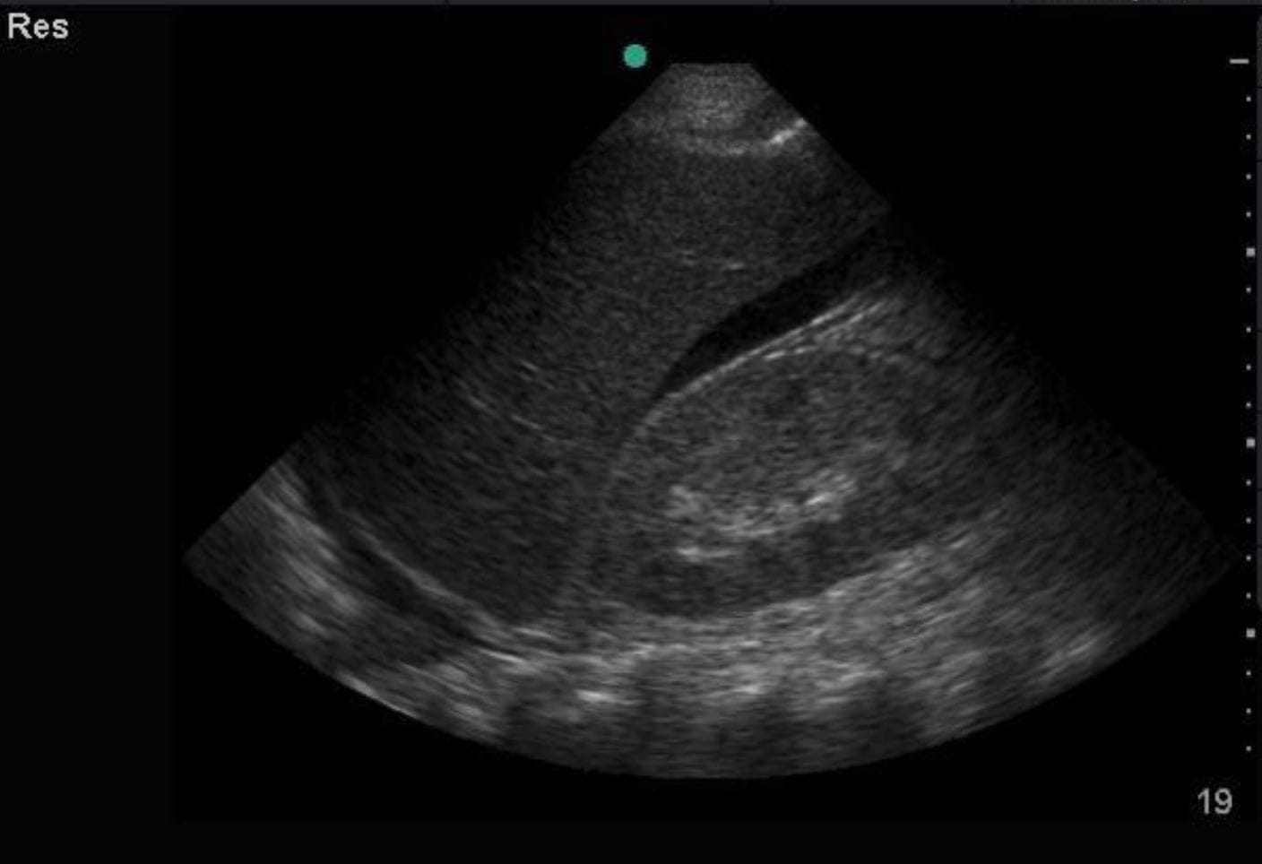
PV Card: FAST Ultrasound
Keeping in line with the recent ultrasound posts this month, Dr. Mike Stone's star team is releasing a series of Paucis Verbis cards on the basics of bedside ultrasonography. Here is the first in the series on the Focused Assessment with Sonography in Trauma (FAST) by Drs. Wilma Chan, John Eicken, and Mike Stone. PV Card: FAST Ultrasound Go to ALiEM (PV) Cards for more resources.
US4TW Case: 28F with Shortness of Breath
Welcome to the inaugural post for an exciting new ultrasound-based case series called “Ultrasound For The Win!” (#US4TW). In this peer-reviewed case series, we will focus on real clinical cases where bedside ultrasound changed the management or aided in the diagnosis. In our first case, we present a 28-year-old female with shortness of breath. [+]

Small bowel obstruction: Diagnosis by ultrasonography
A 64 year old man with an extensive history of abdominal surgeries presents to the emergency department with abdominal pain and vomiting. Because you suspect a bowel obstruction, you bring an ultrasound machine to the bedside prior to the completion of any laboratory testing or other imaging. A curvilinear probe in the abdominal mode setting was used to scan in all four quadrants of the abdomen looking in both the sagittal and transverse planes. [+]

Beta Blockers vs Calcium Channel Blockers for Atrial Fibrillation Rate Control: Thinking Beyond the ED
Intravenous beta blockers and non-dihydropyridine calcium channel blockers are recommended first-line for atrial fibrillation (AF) with rapid ventricular rate (RVR) [1]. In a previous post, Bryan Hayes (@PharmERToxGuy) provided an overview of the data comparing beta blockers to calcium channel blockers for atrial fibrillation rate control in the ED. Here is part 2 of our two-part AF series. [+]

I am giving prochlorperazine. Should I give diphenhydramine too?
Prochlorperazine is a commonly used medication in EM. In certain patients prochlorperazine does wonders for migraines, and remains a great antiemetic choice for undifferentiated nausea/vomiting when ondansetron is ineffective. However, prochlorperazine has antidopinamergic activity increasing the chances of extrapyramidal symptoms (EPS), such as akathisia, dystonia, parkinsonism, and rarely tardive dyskinesia. A common practice in the ED is to give diphenhydramine with prochlorperazine to attenuate EPS. Does this really work? What is the evidence? [+]

Director of Growth, Strategic Alliance Initiative, Center for Innovation and Digital Health
Massachusetts General Hospital;
Chief Scientific Officer, Conductscience.com
Latest posts by Shuhan He, MD (see all)
- The Discharge Severity Index: Early Research on ED Readmission Risk Assessment - August 14, 2025
- Suboxone and the Emergency Physician: Get Waivered Training - August 12, 2022
- A Tale of Two Epidemics: COVID-19 and the Opioid Crisis - April 23, 2020